


ORIGINAL ARTICLES
Background. Scleral rigid gas-permeable contact lenses (SCL) are the preferred method for visual correction in keratoconus (KC). However, complications associated with long-term scleral lens wear remain understudied in the scientific literature. Evaluating the incidence and spectrum of these complications is essential for optimizing patient management and enhancing the safety of scleral lens use in clinical practice. Purpose: To assess the frequency and types of complications associated with long-term scleral lens wear in patients with keratoconus. Materials and methods. The study included 50 patients (100 eyes) with bilateral stage I–IV keratoconus (Amsler–Krumeich classification). The mean age was 29.2 ± 4.3 years. All patients underwent standard ophthalmic examination prior to the selection of optical correction. Slit-lamp biomicroscopy and anterior segment optical coherence tomography (AS-OCT) were used to monitor and identify potential complications during scleral lens wear. Follow-up duration was 12 months. Results. A total of 86% of patients (n = 43) successfully adapted to scleral lenses, with a mean adaptation period of 20.4 ± 5.8 days and an average daily wear time of 11.2 ± 2.4 hours. The primary reasons for discontinuation (14%, n = 7) were handling difficulties (42.9%) and lens damage (28.6%). Among patients who continued lens wear, handling challenges were reported in 25.6%, conjunctival hyperemia in 16.3%, and vision blurring in 9.3%. At the 12-month follow-up, the following complications were observed: mechanical (conjunctival hyperemia – 16.3%, corneal epithelial abrasions – 2.3%), toxic-allergic (2.3%), and infectious (2.3%). No statistically significant correlation was found between daily wear time and complication rate (p = 0.371). However, prolonged adaptation (>20 days) significantly increased the risk of discontinuation (p = 0.002). Conclusion. Long-term scleral lens wear in keratoconus is associated with complications, primarily of mechanical origin. These findings highlight the need for regular follow-up to ensure timely detection and management of complications.
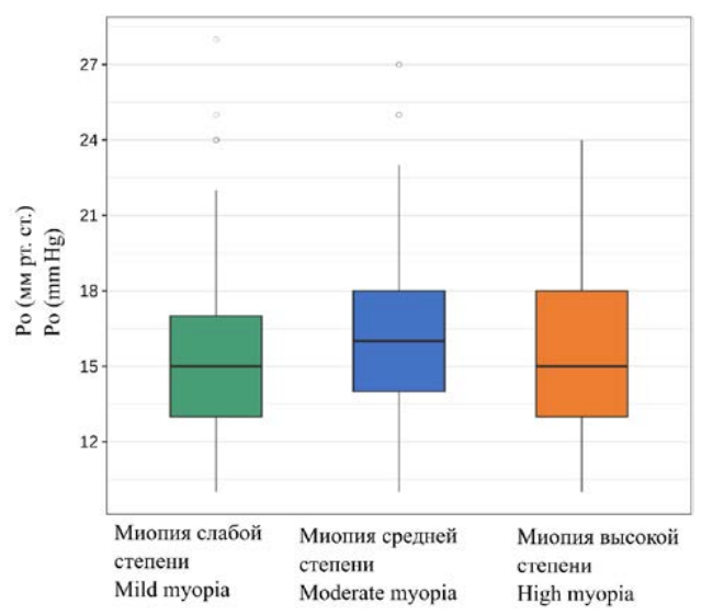
Background. In individuals with myopic refractive error—particularly high myopia—ocular anatomy undergoes changes, most notably an increase in axial length (AL) exceeding 24.5 mm. Several studies have reported alterations in the scleral shell among patients with varying degrees of myopia. Increased myopia is associated with reduced corneal hysteresis and decreased acoustic density of the sclera. Differences in intraocular pressure (IOP) between individuals with myopia, emmetropia, and hyperopia have also been described. However, the available literature provides no conclusive evidence on the relationship between intraocular pressure (IOP) and central corneal thickness (CCT) across different degrees of myopia. Purpose: To assess how intraocular pressure and central corneal thickness vary across different degrees of myopia. Materials and methods. We retrospectively analyzed medical records of 500 patients (1000 eyes) aged 18 years and older, all diagnosed with myopic refractive error. Patients were stratified into three groups based on myopia severity. The cohort included 290 females (58%) and 210 males (42%). Pachymetry was performed using the ALLEGRO Oculyzer (WaveLight Oculyzer II), autorefractometry using TONOREF (Nidek II), and IOP was measured with four tonometers: a non-contact tonometer (TONOREF Nidek II), Maklakov tonometer, Schiøtz tonometer, and Goldmann applanation tonometer. Statistical analysis was conducted using R software (version 4.2.2). Results. A statistically significant positive correlation was observed between increasing myopia and IOP measurements obtained using the non-contact tonometer TONOREF (Nidek II) (p = 0.001) and the Goldmann applanation tonometer (p < 0.001). No such association was found for IOP values measured with the Maklakov (p = 0.978) or Schiøtz (p = 0.262) tonometers. The analysis revealed a statistically non-significant (p = 0.065) trend toward increased central corneal thickness with greater myopia. Mean CCT values were 539.8 ± 32.5 µm in low myopia, 544.1 ± 33.9 µm in moderate myopia, and 546.5 ± 37.9 µm in high myopia. Conclusions. No statistically significant association was found between central corneal thickness (CCT) and the degree of myopia (p = 0.065). The effect of myopia severity on intraocular pressure (IOP) measurements varied depending on the tonometry method used. Statistically significant associations between IOP and myopia severity were observed when measured using the TONOREF (Nidek II) noncontact tonometer (p = 0.001) and the Goldmann applanation tonometer (p < 0.001). In contrast, no such association was found with the Maklakov (p = 0.978) or Schiøtz (p = 0.262) tonometers.
Background. Myopia is the most common refractive disorder of the eye globally. One of the methods for surgical correction of myopia is ReLEx® SMILE (Small Incision Lenticule Extraction). The impact of baseline myopia severity on postoperative outcomes remains debated due to inconsistent literature findings. Purpose: To assess the association between the outcomes of ReLEx® SMILE and baseline myopia severity. Materials and methods. This prospective study included 78 patients (156 eyes) aged 18 to 35 years who underwent refractive correction using the ReLEx® SMILE procedure. Patients were stratified into three groups by myopia severity: Group I – low myopia (n = 31 eyes), Group II – moderate myopia (n = 87 eyes), Group III – high myopia (n = 38 eyes). The follow-up period was six months. Results. Uncorrected visual acuity (UCVA) of 0.9–1.0 was achieved in 100% (Group I), 93% (Group II), and 82% (Group III) of eyes, with statistically significant differences between the groups (p < 0.017). No statistically significant differences in the efficacy index were observed (p = 0.78): efficacy was 100% in Groups I and II, and 97.1% in Group III. The safety index was 100% in Groups I and III and 98% in Group II, with no statistically significant difference (p = 0.458). Refractive regression occurred in one case each in Groups I and III, while no cases of regression were observed in Group II. Conclusion. Refractive correction using the ReLEx® SMILE technique provides predictable and stable correction for all degrees of myopia and myopic astigmatism, with no significant differences in complication rates across severity groups.
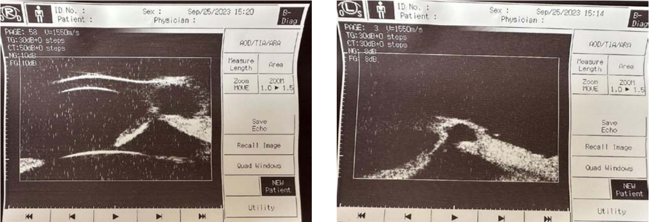
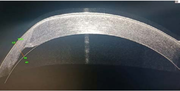
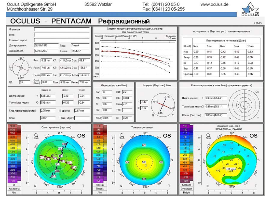
Background. Terrien’s marginal degeneration (TMD) is a rare corneal disorder. Accurate diagnosis is essential but often challenging due to its highly variable clinical presentation. Purpose: to present a clinical case of Terrien’s marginal degeneration. Case description. A 49-year-old male presented to the ophthalmology department at Perm Regional Clinical Hospital in August 2023 with complaints of growths on both eyes. Slit-lamp biomicroscopy revealed marked thinning of the inferior peripheral cornea in both eyes, accompanied by stromal scarring and conjunctival vascular ingrowth, forming a pseudopterygium. Autorefractometry showed against-the-rule astigmatism in both eyes. Anterior segment OCT of both eyes revealed a cavitary structure bounded by thinned corneal stroma and adjacent iris tissue. displaced anteriorly toward the cornea with the formation of iridocorneal synechiae in the anterior chamber angle bilaterally. Ultrasound biomicroscopy confirmed these findings. Based on the clinical history, comprehensive ophthalmologic examination, and anterior segment OCT data, a diagnosis of Terrien’s marginal degeneration complicated by corneal staphyloma was established. Conclusion. This clinical case of Terrien’s marginal degeneration in a 49-year-old male underscores the importance of accurate diagnosis. The disease course and clinical features are consistent with the limited published descriptions of this condition. Ancillary imaging techniques, including anterior segment OCT and ultrasound biomicroscopy, enhance diagnostic accuracy in atypical presentations. Patients with TMD require long-term ophthalmic follow-up.
Background. Selective keratoplasty techniques, in which only the affected corneal layers are replaced, are increasingly used in modern corneal transplantation. In penetrating keratoplasty (PK), however, all layers of a structurally altered cornea are replaced with a donor graft. PK remains the only surgical option for many severe anterior segment conditions, including advanced keratoconus, corneal ulcer with stromal melting and/or perforation, and corneal opacities of various origins involving the full corneal thickness. Purpose: to report clinical cases of penetrating corneal grafts with transparency preserved for more than 20 years. Materials and methods. In 2024, 19 patients (24 eyes) with penetrating corneal grafts performed by the authors more than 20 years earlier (20– 35 years) were examined. The cohort included 10 men and 9 women. In all cases, fresh (non-stored) donor corneas were used, with a death-to-surgery interval of less than 24 hours. The graft diameter was ≥8 mm in all eyes. The primary diagnosis was advanced keratoconus (stage IV by Amsler, or post-hydrops scarring) in 13 patients. The remaining six patients had dense corneal scars and opacities due to penetrating trauma, severe keratitis, or ulcers (including cases with perforation). Results. In all 24 eyes, the corneal grafts remained transparent; two cases showed progressive endothelial graft dysfunction. Five representative cases of ultra-longterm graft survival and one case of bilateral reconstructive PK performed in Minsk, Republic of Belarus (1 and 8 years post-surgery), are illustrated. Conclusion. Penetrating keratoplasty with fresh donor corneas of high endothelial cell density provides the potential for successful graft survival with long-term–and even ultra-long-term–maintenance of transparency. In keratoconus, refractive PK may result in lower post-keratoplasty ametropia, better visual function, and a reduced risk of ectasia recurrence.
Background. Modern cataract surgery requires increasingly high standards to achieve excellent visual outcomes at all distances, with mandatory intraoperative correction of corneal astigmatism. In such cases, customized intraocular lenses (IOLs), calculated according to the patient’s specific biometric parameters, are used. This report presents a clinical case of implanting a customized toric multifocal IOL during cataract surgery in a patient with high axial myopia and concomitant corneal astigmatism. Case description. A 45-year-old male with complicated cataract, high axial myopia, and corneal astigmatism underwent phacoemulsification with implantation of a customized multifocal IOL (Lentis Mplus LU-313T). In addition to standard ophthalmological examinations, optical specular endothelial microscopy of the cornea, optical coherence tomography (OCT) of the retina, and Scheimpflug corneal densitometry were performed. Preoperative IOL power calculation was carried out using an online calculator based on IOLMaster and corneal topography data. Postoperatively, uncorrected visual acuity was 1.0 for distance, 0.4 at intermediate, and 0.5 at near. Discussion. Implantation of customized IOLs with rotationally asymmetric optics, with a +2.0 D addition in the dominant right eye (Lentis Mplus LU-313 MF-20T) and a +3.0 D addition in the nondominant left eye (Lentis Mplus LU-313 MF-30TX), eliminated the need for postoperative optical correction at different distances. High uncorrected visual acuity was achieved for distance, intermediate, and near vision. The absence of halo effect after surgery enabled the patient to drive comfortably, including in the evening and at night. Conclusion. This comprehensive surgical approach to treating complicated cataract in eyes with high myopia and corneal astigmatism provided complete visual rehabilitation and eliminated the need for additional optical correction.
REVIEWS
Background. Glaucoma is a chronic progressive disease leading to significant vision loss and remains one of the leading causes of blindness worldwide. Among the most effective treatment options is surgical intervention aimed at lowering intraocular pressure (IOP) by creating or restoring aqueous humor outflow pathways. However, the major challenge of glaucoma surgery is postoperative fibrosis, which reduces surgical success. Purpose. To systematize current understanding of the wound-healing response following glaucoma surgery and to identify key strategies for controlling scarring. Materials and methods. The first part of the review includes scientific articles published over the past 10 years, focusing on wound-healing mechanisms in glaucoma surgery, methods for preventing fibrotic complications, and promising therapeutic strategies. A literature search was conducted in PubMed, Scopus, and Web of Science databases, yielding 59 articles for analysis. Results. The analysis showed that the main drivers of postoperative fibrosis after glaucoma surgery are the activation of inflammatory pathways, overproduction of extracellular matrix, and immune dysregulation. Antimetabolites remain the gold standard for preventing postoperative fibrosis. Mitomycin-C (MMC) significantly improves trabeculectomy outcomes, but at high doses it may induce dystrophic and necrotic changes in ocular surface tissues. By contrast, 5-fluorouracil (5-FU) has a safer profile but requires serial administration and is less effective. Combining low doses of antimetabolites with an amniotic membrane or hydrogel carriers reduces toxicity and prolongs drug action. Targeted inhibition of growth factors—including angiogenesis inhibitors, pirfenidone, and SB-431542—demonstrates high therapeutic potential. Conclusion. Effective management of reparative processes after glaucoma surgery requires a multi-tiered approach, encompassing inflammation reduction, modulation of cellular signaling pathways, and controlled extracellular matrix remodeling. Despite progress, further research is needed to optimize combination therapies, minimize adverse effects, and implement advanced biotechnological strategies for greater precision in clinical practice.
Background. Cytomegalovirus (CMV) retinitis is an inflammatory–necrotic disease that can lead to irreversible blindness. The cornerstone of diagnosis remains ophthalmoscopy. However, ophthalmoscopy is limited to direct visualization of retinal and choroidal changes, whereas optical coherence tomography (OCT) provides insight into structural alterations and spatial relationships within the vitreoretinochoroidal complex, thereby offering new diagnostic opportunities. Purpose: To systematize available data on the manifestations of CMV retinitis as assessed by OCT. Materials and methods. Publications were searched in PubMed, Google Scholar, Web of Science, and Scopus using the following keywords: “optical coherence tomography”, and “cytomegalovirus retinitis” (both in English and Russian); “retinal necrosis”, “ocular involvement in immunodeficiency”, “OCT angiography”, and “vitreoretinal interface” (in Russian only). Thirty publications describing the clinical features and case reports of CMV retinitis with OCT findings were selected. Results. This review summarizes distinguishing features between acute and resolved inflammation, differences in retinal perfusion in this patient group based on OCT angiography, and several hypotheses concerning the entry and intraocular dissemination of cytomegalovirus infection. Publications were structured according to OCT capabilities and study design, ranging from retrospective analyses of prior retinonecrosis cases to prospective investigations and disease monitoring using advanced OCT angiography techniques. Conclusion. OCT enables differentiation between active and inactive lesions of CMV retinitis and allows preclinical visualization of CMV-related changes within the eye.
WORKSHOP
The introduction of oxygen-permeable rigid gas permeable (RGP) lenses marked a significant turning point in the history of contact lens wear. Prior to their development in the late 1970s, polymethyl methacrylate (PMMA) lenses dominated clinical practice. PMMA is entirely impermeable to oxygen, and while its optical clarity and mechanical durability were unmatched for decades, the physiological burden placed on the cornea by prolonged wear of these lenses became increasingly evident. Chronic corneal hypoxia was an inevitable consequence of PMMA lens wear, leading to a series of metabolic, structural, and functional changes in the cornea that persist long after lens removal.
When transitioning a long-term PMMA wearer to a modern high Dk RGP material, the clinician must not only consider the optics and fit but also the underlying metabolic state of the cornea. Understanding the cellular and biochemical consequences of hypoxia—and the challenges associated with sudden reoxygenation—is essential to ensuring a successful and comfortable adaptation to new lens materials.
TECHNOLOGIES
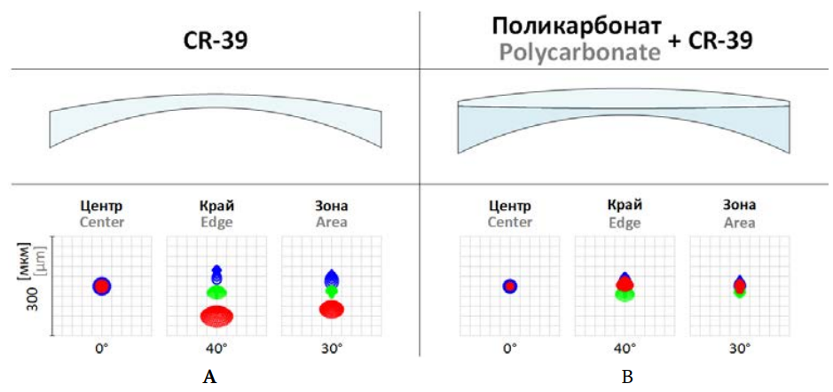
This study proposes an approach based on the use of a special adhesive composition for cementing polymer lenses, enabling the creation of multilayer optical systems for corrective glasses with more effective aberration correction.
TECHNOLOGIES IN EDUCATION
Objective. To define the role and place of graduate school (aspirantura) in the system of training scientific and pedagogical personnel, revealing its essence as a synthesis of the educational process and research activity, as well as to assess its impact on the professional trajectories of graduates. Methods. The study employs a systemic-structural analysis, which allowed for the consideration of graduate school as a complex institution that combines the educational process and research activity. Results. The key functions of graduate school are identified: ensuring the continuity of scientific traditions, developing the country’s scientific potential, and forming a personnel reserve for academia and research institutions. The organizational stages (admission, candidate exams, research work, dissertation defense) are detailed, and its substantive specificity is revealed, which differs from previous levels of higher education by its emphasis on the production of new knowledge. It is established that obtaining a Candidate of Sciences degree expands opportunities for professional fulfillment both within the academic sphere (universities, research institutes) and beyond (analytical centers, high-tech companies). Conclusions. Graduate school represents a unique social elevator and a crucial institution that shapes specialists capable of independent scientific creativity, analytical work, and solving complex professional tasks. Despite its high complexity, it provides unique research experience that remains a foundation for an entire subsequent career.


