


EDITORIAL

ORIGINAL ARTICLES

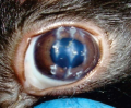
Background. Keratoconus (KC) is the most commonly diagnosed corneal ectasia among young adults today. The asymmetric process of progression of the KС determines the occurrence of anisometropia, which in a compartment with increased audience and the absence of proper optical correction leads to a violation of the accommodation function that has not previously studied with this pathology.
Purpose: to study the work of accommodation using an objective method of computer accommodation in young people with newly diagnosed keratoconus (КC).
Material and methods. Thirty (60 eyes) people were included in the study, of which 15 people (30 eyes) formed the main group and 15 (30 eyes) formed the control group. The main group consisted of patients with newly diagnosed bilateral KC: 15 eyes – stage 1, 13 eyes – stage 2, 2 eyes – stage 3 of the disease). Patients of the control group had a history of mild myopic astigmatism of the direct type. The average age of the patients was 23.5 ± 3.1 years. All patients underwent standard ophthalmological examination: autorefkeratometry, visometry and biomicroscopy. In addition, the main keratometric parameters of the cornea were examined using the Scheimpflug analyzer Pentacam. An objective assessment of the indicators of the accommodative ability of the eye was carried out using computer accommodation on the Righton Speedy-I device. The statistical processing was carried out in the IBM SPSS Statistics 23 program.
Results. The results of the analysis of accommodograms in both groups demonstrate the predominance of disorders of the ciliary muscle, which manifested themselves in the form of habitual-excessive accommodation tension (HEAT) and accommodative asthenopia of a spasmodic nature. In the control group, in 76% of cases, there was a pattern of excessive accommodative response by the type of HEAT. The state of accommodation in the group with KC in 68% of cases was characterized by an excessive and unbalanced accommodative response, with an unstable course of the curve. The average indicator and the curve of the accommodogram significantly exceeded the indicators of the control group, which proves an unstable state of accommodation in KC. In comparison with the control group, a decrease in coefficient of growth of the accommodogram (CR) by 37% (p < 0.05) was recorded, confirming the absence of a smooth and increasing course of the accommodative response following the stimulus. Analysis of high-frequency the coefficient of microfluctuation (CMF) demonstrated a 10% increase in the work of the ciliary muscle (p < 0.001). The correlative dependence of these parameters with visometry and autorefractometry data has been established.
Conclusion. The objective method of accommodation assessment revealed abnormalities of ciliary muscle functioning in patients with a first-time diagnosis of KC, which correspond to the phenomena of spasmodic accommodative asthenopia. The established correlation between CR and CMF with the data of visometry and autorefractometry suggests a potential improvement of accommodation functioning under conditions of optical correction with contact lenses.

Glaucoma continues to lead among the irreversible causes of blindness and low vision. In some cases, surgical treatment is the method of choice even for newly detected glaucoma. The indication for the use of drains is refractory glaucoma, but today microshunting is widely enough used in patients with primary open angle glaucoma at the first stage of surgical treatment.
The aim of the study is to develop a technology of polymeric microshunt implantation and estimate the clinical effect of its using for the surgical treatment of worsening and terminal open‑angle glaucoma.
Materials and Methods. 30 patients (30 eyes) with open‑angle glaucoma and high level of intraocular pressure (IOP) were observed. All the patients were given modified partial‑thickness deep sclerectomy with the polymeric microshunt implantation by means of the developed insertor. The shunt made of polymer (polycarbonate methacrylate) is a hollow square tube 2.5 mm long, the diameter of the inner hole is 0.2 mm. The needle cut angle is 45 degrees, the end of the needle has an additional lateral anti‑lock hole. The microshunt consists of two parts: the needle for placing into the anterior chamber and supporting elements for the shunt fixation between the superficial and deep layers of the sclera. The insertor we created is an instrument 12.2 mm long with the 2.2 mm long thin rod at the end. Before implantation the shunt put on the needle is securely fixed due to the working part of the tool cut (45 degrees), which is congruent to the external part of the shunt. After the implantation the needle is removed from the shunt by pressing the side button of the insertor that allows to leave the shunt at the implantation site.
Results. All the patients demonstrated good results at a distant time with areactive current of postoperative period. Computer static projector perimetry and visometry didn’t reveal any negative dynamics during the entire observation period. According to ophthalmoscopy and optical coherent tomography results, the changes of the optic disc, ganglion cells layer in the macula and peripapillary nerve fibers remained at the same level.
Conclusion. Antiglaucomatous surgery according to the invented technology of the polymeric shunt implantation with the help of the original insertor in combination with modified sclerectomy provides stable hypotensive effect and minimizes complications. In addition, the method helps to stabilize visual functions in patients with worsening and terminal open‑angle glaucoma and high level of IOP.

Background. Endolaser coagulation (ELС) is an integral part of endovitreal retinal detachment (RD) surgery. The study of the role of ELC volume (total laser energy and the amount of applied coagulates) in the formation of perisilicone oil proliferation (POP) after RD surgery of various origins is necessary to prevent postoperative complications.
The purpose of this study was to study the role of the ELC volume in the formation of POP in RD surgery of various genesis.
Materials and methods. The study was conducted in the private clinic “SIHAT KO’Z” from 2019 to 2021. 88 (88 eyes) patients with retinal detachment of different genesis were examined. The age range varied from 21 to 67 years, the average age was 47.4 ± 3.1 years. 61 (69%) patients had rhegmatogenous RD, 27 (31%) tractional RD. The macular area was involved in RD in 64 (73%) cases, and was intact in 24 (27%) cases.
Results. In 11 patients PP formed the day after surgery, in 4 (4.5%) exudate and fibrin were formed in the anterior chamber, in 4 (4.5%) re‑injection of silicone oil was required due to incomplete retinal adhesion. In 2 (2.3%) cases iridocorneal adhesions were formed. The study showed that the occurrence of the above complications was directly related to the intensive effect of ELC during surgery for retinal detachment.
Conclusion. Thus, increased coagulation and energy volume in ELC during surgical treatment of retinal detachment is important in the occurrence of PP. The analysis revealed a clear relationship between the development of a proliferative membrane and the volume of ELC. Since PP is a factor that reduces the effectiveness of surgical treatment of retinal detachment, the recommendation is to reduce the volume of ELC to the minimum necessary.

Most ophthalmic interventions are examples of microinvasive surgery using microscopes of various modifications, which can have a negative impact on the eye condition of ophthalmic surgeons. Thus, presumably, a prolonged and repeated change in the accommodation of surgeons can contribute to a change in the dynamic refraction of the eye; separate articles began to appear describing the development of artifical dry eye syndrome (DES). It seems obvious that systematic work in the operating room and the conduct of microsurgical operations affect the organ of vision of ophthalmic surgeons.
Purpose: to determine the degree of influence of equipment intended for microsurgical ophthalmic operations on the dynamic characteristics of the level of intraocular pressure, refraction and the state of the eye surface of ophthalmic surgeons.
Materials and Methods. As part of a multicenter analytical scientific cross‑sectional study, data from 48 people (48 eyes, men – 50% and women – 50%) were analyzed. The indicators of total tear production (Schirmer test, I), as well as individual tonometry and refractive index characteristics of volunteers at the beginning and at the end of the surgical day, were studied. Individual risk factors for the development and progression of the dry eye syndrome (age, smoking, use of hormonal systemic drugs, wearing soft contact lenses), as well as instillation of artificial tears, were recorded. Additionally, all subjects completed the Ocular Surface Disease Index (OSDI) questionnaire.
Results. After the operation day, which averaged 2.8 ± 0.2 hours, the subjects (mean age – 41.9 ± 1.1 years) had a statistically significant change in the index of tear production (from 11.9 ± 0.9 to 10.8 ± 1.7 mm, p = 0.01; Z = 2.407). A significant decrease in the parameters of the Schirmer test 1 was found in the age group of surgeons under 40 years old (p = 0.018; Z = 2.353), and in persons over 40 years old, there was a tendency for a decrease in tear production parameters, which was not statistically significant (p = 0.213; Z = 1.244). There was a tendency to increase the IOP‑level in surgeons of both age groups (p = 0.314; Z = 1.006 and p = 0.632; Z = 0.407, respectively). In addition, in surgeons older than 40 years, there was a change in accommodation, expressed in an increase in myopic refraction by 0.5 diopters at the end of the surgical day (p = 0.076; Z = 1.771).
Conclusions. The use of specialized professional equipment (operating microscope) was found to have a negative effect on total tear production regardless of the presence of traditional risk factors for DES. The severity of violations depended on the age category of the subjects. The data obtained are of particular relevance in the period of development and progression of methods of surgical treatment of ophthalmic diseases and substantiate the need to develop specific preventive measures.
The evolution of therapeutic technologies dictates the development of a strategy for their implementation in clinical practice. The first stage is the assessment of the capabilities, efficiency, advantages and disadvantages, development of indications and contraindications on models of eye diseases in the experiment.
The purpose of the work: to develop criteria for assessing the effectiveness of anti‑angiogenic drugs and evaluate the proposed modeland neovascularization of the eye in the experiment.
Materials and methods. Neoangiogenesis models – two chorioretinal and two corneal localizations – were formed in the experiment on rabbits of Sovetskaya Chinchilla breed. The advantages and disadvantages of each of them were analyzed empirically. Experimental animals with neovascular disease were treated with recombinant peptides with angiostatic potential (tumstatin, PEDF, endostatin). In the course of treatment, we developed efficacy criteria based on clinical, instrumental, and morphological studies.
Results. Comprehensive efficacy evaluation criteria were developed and tested, allowing not only to reveal and describe the spectrum of biological effects of the tested drugs, but also to quantitatively assess and compare their therapeutic potential with each other at almost all stages of vascular assembly. The complex consisted of qualitative and quantitative clinical (hyperemia, edema, vascularization, antiangiogenic activity according to a score scale), angiographic (intensity and area of edema, area of neovascularization and index of corneal vascularization) and morphological (extent of edema, extent of vessels and their number in section, index of vascularization) indicators.
Conclusion. To get the full amount of information about the tested drug with the claimed angiostatic potential, it is necessary to use several models of neoangiogenesis with different mechanisms to study the amplitude and spectrum of their effects.
WORKSHOP

Presbyopia is becoming a global problem worldwide, leading to reduced quality of life due to lack of correction or hypocorrection in 94% of people over 45. Multifocal soft contact lenses (MFCLs) are one of the fastest growing methods of contact correction for presbyopia. According to a report by our international colleagues, MFCLs represent 14% of all types of MCLs worldwide in 2021, an increase of 2% from 2020.
In this article we will look at the OKVision® Fusion NEW Multifocal fitting algorithm to help professionals simplify and speed up the fitting process and help patients get the vision quality they want at the right distances.
DISCUSSION CLUB
LITERARE GUIDE
MEDICINE AND LAW
Purpose: to share our experience of regulatory justification to supervisory authorities of performing keratoplastic operations using native donor cornea.
Methods. A comprehensive analysis of legal and regulatory framework of the Russian Federation on transplantation.
Results. The Nomenclature of Medical Services includes several types of services which contain the word “transplantation” in their names. However, not all of them require a license to perform such procedures as “surgery (organ and (or) tissue transplantation)”. The procedure for providing medical care in “surgery (transplantation of organs and (or) human tissues)” is not applicable to the “ophthalmology” profile, since the equipment and staffing requirements contained therein are not necessary to perform keratoplasty. The list of medical organizations of the subjects of the Russian Federation performing transplantation of human organs and (or) tissues contains no medical organizations of the ophthalmological profile. According to methodological recommendations on ways to pay for medical care at the expense of mandatory medical insurance, keratoplasty is classified as “st21.005. Operations on the organ of vision”.
Conclusion. A comprehensive analysis of existing regulatory documents demonstrates that keratoplasty is exclusively related to ophthalmology, it can be performed if there is a license for ophthalmology, transportation of human organs and (or) tissues for transplantation. A healthcare organization also needs to hold a license for removal and storage of human organs and (or) tissues for transplantation. However, if any healthcare organization in the region has such a license, it may donate a donor cornea to an ophthalmological hospital if the latter does not hold such a license.
NEWS: WHAT? WHERE? WHEN?


