Open Access
 Subscription or Fee Access
ORIGINAL ARTICLES
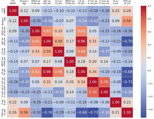
Background. Myopia remains one of the leading causes of visual impairment worldwide. A wide range of therapeutic and preventive strategies is currently applied in clinical practice to slow myopia progression. In recent years, increasing attention has been directed toward the use of artificial intelligence (AI) for predicting disease progression and treatment outcomes. Machine learning techniques enable the development of predictive models based on baseline clinical data, thereby improving individualized treatment planning. Purpose: To develop and validate a machine learning model for predicting myopia progression at the 12-month follow-up in children wearing peripheral defocus spectacle lenses. Materials and methods. A dataset comprising 48 eyes of 48 pediatric patients fitted with peripheral defocus spectacle lenses was analyzed. Binary classification models were developed using Python 3 and the scikit-learn, XGBoost, and LightGBM libraries. Eight machine learning algorithms were evaluated: XGBoost, Random Forest, Gradient Boosting, LightGBM, Extra Trees, Logistic Regression, Decision Tree, and K-Nearest Neighbors. The outcome variable was defined as a binary indicator of treatment effectiveness: favorable outcome (24 eyes) – annual myopia progression less than −1.00 diopters at 12 months; unfavorable outcome (24 eyes) – progression of −1.00 diopters or greater. The dataset was divided into training and test sets in a 67:33 ratio. Feature importance was assessed using built-in feature importance methods. Results. The highest predictive performance on the test dataset was demonstrated by the XGBoost model, with a ROC AUC of 0.906. The model achieved an accuracy of 0.875, sensitivity of 0.875, and specificity of 0.875. The most influential predictors of treatment outcome were: minimum keratometry (Kmin) at baseline (0.382), baseline spherical equivalent refraction (0.217), baseline corneal radius of curvature (0.184), baseline axial length (0.102), patient age (0.064), and maximum keratometry (Kmax) at baseline (0.051). Conclusion. The proposed machine learning model demonstrated excellent predictive performance for forecasting the outcome of peripheral defocus spectacle lens wear in children at the 12-month follow-up. A clinical decision-support calculator based on this model has been developed for practical application.
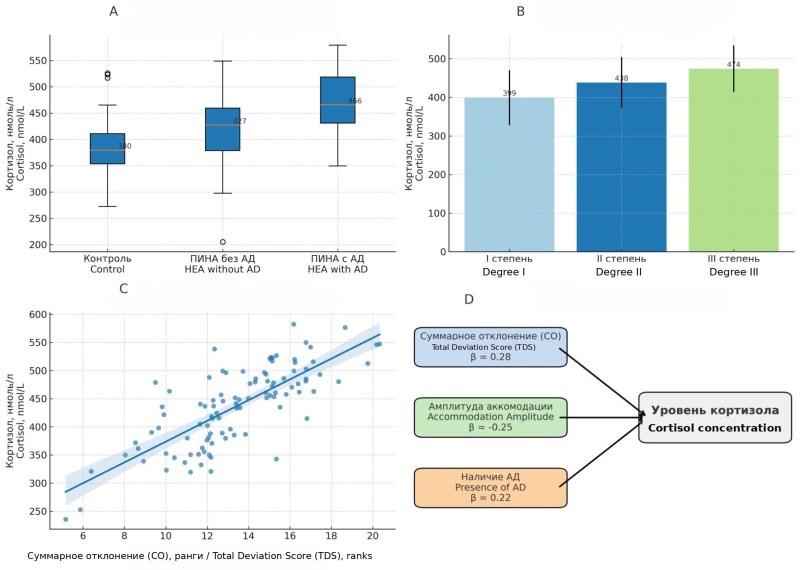
Background. Habitual excessive accommodation (HEA) in children represents a multifactorial pathophysiological condition involving ophthalmic dysfunction alongside autonomic and neuroendocrine dysregulation. Identification of pathogenetic interrelations is essential for the development of integrated diagnostic and therapeutic approaches aimed at preventing progression of both visual and systemic disturbances. Purpose: To assess the psychoemotional status of children with habitual excessive accommodation and to examine its relationship with clinical manifestations of accommodative dysfunction. Materials and methods. Between May and July 2025, 150 children aged 6–17 years were examined at City Clinical Polyclinic № 29, including a main group with habitual excessive accommodation (n = 102) and a control group (n = 48), comparable in age and sex. Ophthalmic examination included assessment of uncorrected and best-corrected visual acuity; manifest and cycloplegic refraction; and accommodative amplitude, reserve, and facility. Psychoemotional status was evaluated using the eight-color Lüscher test with calculation of the autonomic coefficient (AC) and total deviation score (TDS). Serum cortisol concentration was measured in the morning under fasting conditions by enzyme-linked immunosorbent assay (ELISA). Results. Children with HEA showed a statistically significant reduction in accommodative amplitude (8.6 ± 1.9 D vs 11.2 ± 2.1 D in controls), accommodative reserve (1.4 ± 0.6 D vs 2.6 ± 0.7 D), and accommodative facility (5.3 ± 1.8 vs 9.1 ± 2.0 cycles/min). Accommodative parameters decreased with increasing HEA severity. Psychoemotional assessment revealed a higher median TDS (14 ranks vs 8 in controls) and elevated AC values (1.6 ± 0.8 vs 0.4 ± 0.7), reflecting sympathetic predominance. Autonomic dissociation (AC > +1.0 combined with altered color preference patterns) was identified in 38.2 % of children with HEA and was associated with lower accommodative parameters and higher TDS values (median 16 ranks). Serum cortisol levels exceeded those of the control group (432 ± 70 vs 368 ± 65 nmol/L), particularly in children with autonomic dissociation (456 ± 68 nmol/L). Cortisol levels correlated positively with TDS (ρ = 0.47) and negatively with accommodative amplitude (ρ = −0.41). Conclusion. The study demonstrated that habitual excessive accommodation in children is characterized by a combined ophthalmic, psychoemotional, and neuroendocrine imbalance. A high level of psychoemotional stress was recorded, associated with autonomic dissociation and a shift of autonomic balance toward sympathetic predominance; increased basal cortisol levels reflect systemic involvement of stress-realizing mechanisms. This indicates disintegration of autonomic nervous system regulators and supports the need for a comprehensive approach to correction.
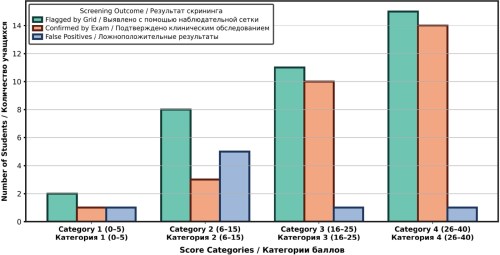
Background. Children’s visual health represents a major public health concern, particularly in low- and middle-income countries where access to specialized eye care is limited. In Morocco, the scarcity of eye care professionals poses a significant challenge to implementing large-scale school-based vision screening programs. Objective: this study aimed to evaluate the effectiveness of a low-cost vision screening method – a teacher-administered observational vision screening test – as an alternative approach for the early detection of visual impairment in primary school children. Materials and methods. The study included 143 children aged 6–12 years from two Moroccan primary schools. Teachers assessed vision-related signs and symptoms using a structured behavioral checklist comprising ten predefined indicators. Each child received a cumulative score, which was used to determine the level of risk for visual impairment and the need for referral. All participants subsequently underwent a comprehensive ophthalmological examination performed by an ophthalmologist and an orthoptist. Diagnostic accuracy was assessed using sensitivity, specificity, and predictive values. Results. The screening method identified 36 children as being at risk for visual impairment, of whom 28 were clinically confirmed, corresponding to a prevalence of 19.6 % in the study population. The method demonstrated high diagnostic performance: sensitivity – 100 %, specificity – 93.0 %, positive predictive value (PPV) – 77.8 %, negative predictive value (NPV) – 100 %, and overall accuracy – 94.4 %. Agreement between teacher-based screening results and clinical diagnosis was high (κ = 0.84). Conclusion. The findings indicate that, with minimal training, teachers can effectively identify children at risk of visual impairment using a simple observational screening method without direct involvement of medical personnel. This approach represents a practical, scalable, and cost-effective strategy for school-based vision screening in resource-limited settings.
Introduction. Patients with age-related cataracts frequently present with comorbid conditions such as glaucoma, diabetes mellitus, or a history of uveitis. These conditions may complicate cataract phacoemulsification due to the presence of small, rigid pupils. In such cases, the use of iris retractors is required, which is often associated with intraoperative and postoperative pain. Therefore, anesthesia that ensures adequate analgesia without causing discomfort or additional complications is essential for these surgical interventions. Objective: to identify the most effective anesthetic technique and anesthetic agent in complicated phacoemulsification cases requiring the use of iris retractors. Materials and methods. This study was conducted between May 2016 and May 2018 at the medical center MFC-Kuzbass LLC. A total of 56 patients (56 eyes) with cataract complicated by pseudoexfoliation syndrome (PEX), rigid pupils, and posterior synechiae were included. All patients underwent phacoemulsification with the use of iris retractors. Using a random number generator, patients were allocated into three groups: sub-Tenon’s anesthesia with 0.75 % ropivacaine (n = 18), sub-Tenon’s anesthesia with 2 % lidocaine (n = 19), and topical (epibulbar) anesthesia with 0.5 % proxymetacaine (n = 19). All patients underwent a comprehensive ophthalmic examination and standard preoperative laboratory testing. Pain intensity was assessed using a visual analog scale (VAS) ranging from 1 to 10 points, immediately after surgery and 24 hours postoperatively. Statistical analysis was performed using SPSS Statistics 22.0 with nonparametric tests (Mann– Whitney U test and Kruskal – Wallis test). Pain intensity was expressed as the median and interquartile range. Results. During surgery with iris retractors, the median pain score was 3.5 (2.0–5.0) in patients receiving sub-Tenon’s anesthesia with 0.75 % ropivacaine, 7.5 (6.0–8.0) in those receiving topical anesthesia with 0.5 % proxymetacaine, and 5.0 (5.0–5.0) in patients receiving sub-Tenon’s anesthesia with 2 % lidocaine. Intravenous analgesia during surgery was required in 11 patients from the proxymetacaine group and in 5 patients from the lidocaine group. No patients in the ropivacaine group required additional intravenous analgesia. In the early postoperative period, analgesics were not required in the ropivacaine group, whereas analgesics were prescribed to 6 patients in the proxymetacaine group and to 2 patients in the lidocaine group. Conclusion. Sub-Tenon’s anesthesia with 0.75 % ropivacaine demonstrated the highest effectiveness in complicated phacoemulsification cases involving iris retractors and can therefore be recommended for broader clinical use. Pharmaceutical companies should consider expanding approved indications for anesthetic agents in ophthalmology, including conducting safety studies on their intracameral use.
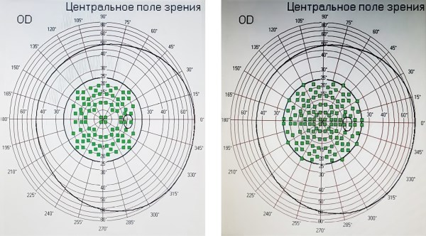
Background. In a substantial proportion of patients with full-thickness macular holes, recurrence or persistence of the defect may occur even after technically successful primary surgery. At present, no universally accepted surgical strategy exists for the management of persistent macular holes. Purpose: to evaluate the clinical effectiveness of a newly developed combined surgical technique based on internal limiting membrane flap reconstruction for macular holes that failed to close after primary surgery. Materials and methods. The outcomes of the proposed surgical technique were analyzed 3–4 years after reoperation for persistent macular holes. The study included 92 patients aged 66.5 ± 5.5 years who underwent repeat surgery for persistent full-thickness macular holes. Two surgical approaches were evaluated: (1) use of a free internal limiting membrane flap combined with gas-air tamponade and application of autologous conditioned plasma (Group 1, n = 41); (2) use of a free internal limiting membrane flap with silicone oil tamponade (Group 2, n = 51). Optical coherence tomography and microperimetry were the primary assessment methods. Results. Both techniques demonstrated high anatomical success rates and resulted in macular hole closure. However, the technique involving a free internal limiting membrane flap with ACP and gas-air tamponade yielded superior functional outcomes. Patients in this group showed greater improvement in visual acuity (by 0.04; p = 0.003) and retinal sensitivity (by 2.15 dB; p = 0.04) compared with Group 2. These differences are presumably related to the absence of toxic and mechanical effects associated with silicone oil tamponade. Conclusion. The proposed technique using a free internal limiting membrane fl ap combined with gas-air tamponade and autologous conditioned plasma is preferable in terms of functional outcomes, complication minimization, and reduced need for repeat surgical interventions.
REVIEWS
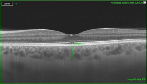
Background. Optical coherence tomography is an indispensable diagnostic tool in ophthalmology, providing high-resolution, non-invasive in vivo visualization of choroidal structural characteristics. Advances in imaging technologies have enabled the identifi cation of new quantitative and qualitative parameters, either calculated by built-in tomograph software or derived using additional image-processing programs. At the same time, the need for unified assessment criteria and systematic classification of existing parameters remains relevant, as this would enhance their clinical applicability and improve the reliability of research data. This review analyzes the capabilities of optical coherence tomography in the evaluation of the choroid. Purpose: To summarize published data on quantitative and qualitative parameters characterizing the choroid as assessed by optical coherence tomography. Materials and methods. A literature search was conducted in the following databases: CyberLeninka, eLIBRARY. ru, PubMed, Embase, Google Scholar, Web of Science, and MEDLINE, covering the period from January to March 2025. A total of 98 publications were identifi ed, of which 61 were included in the review. Both clinical studies and review articles published in Russian and English over various time periods were analyzed. The search keywords included choroid, optical coherence tomography, subfoveal choroidal thickness, choroidal thickness map, choroidal contour mapping, choroidal vascularity index, choroidal vascularity map, choroidal volume, and ImageJ. Results. OCT-based assessment of the choroid includes measurement of subfoveal choroidal thickness, choroidal thickness at specific locations, and the choroidal vascularity index. In addition, next-generation optical coherence tomography software enables evaluation of choroidal features through choroidal thickness maps, choroidal contour mapping, choroidal vascularity maps, and choroidal volume analysis. This comprehensive approach allows a transition from point-based measurements to detailed three-dimensional modeling and functional assessment of the choroid. Conclusion. Investigation of novel structural characteristics of the choroid may contribute to a deeper understanding of the role of the choroid in the pathogenesis of ocular diseases.
Cyclosporine A (CsA) is a neutral, lipophilic, hydrophobic cyclic polypeptide derived from the fungal metabolites Tolypocladium inflatum and Beauveria nevus. Despite the widespread introduction of topical CsA formulations into clinical practice, their use in inflammatory ocular surface disorders and dry eye disease remains largely based on heterogeneous studies and empirical clinical experience, without a unified, systematic appraisal of the available evidence. The absence of a structured review comparing different CsA formulations, their immunomodulatory mechanisms, clinical indications, and limitations represents a significant scientific and clinical gap. Purpose: to systematize data from the scientific literature on topical CsA as a well-established therapeutic agent for inflammatory ocular surface disorders and dry eye disease. Materials and methods. Data from original studies were analyzed, and published findings from the most relevant works on CsA formulations used in ophthalmology were synthesized. Publications were identified using a comprehensive search strategy based on predefined keywords. A total of 93 articles indexed in the PubMed database were included in the review. Results. In ophthalmology, 0.05 % CsA was the first commercially available formulation approved for the treatment of dry eye disease and registered in the Russian Federation. Subsequently, a 0.1 % cationic emulsion was introduced for the same indication. The literature analysis demonstrates that topical CsA increases tear production and exerts a protective effect on the ocular surface epithelium and conjunctival goblet cells. Its therapeutic immunomodulatory action is associated with a reduction in HLA-DR expression, pro-inflammatory cytokine levels, apoptosis markers, and conjunctival T-lymphocyte infiltration. Conclusions. In ophthalmic practice, CsA should be used for a relatively narrow but therapeutically challenging group of ocular surface diseases, either in combination with topical glucocorticosteroids or as monotherapy in a steroid-sparing regimen. In addition to severe immune-mediated forms of dry eye disease, CsA is appropriate for the management of other T-cell-mediated inflammatory ocular surface conditions, including post-keratoplasty graft rejection, vernal and atopic keratoconjunctivitis, keratitis associated with autoimmune diseases, Thygeson’s superfi cial punctate keratitis, and meibomian gland dysfunction.
Background. Isotretinoin remains one of the most effective therapeutic agents for the management of chronic acne vulgaris; however, its use is associated with a wide spectrum of adverse effects varying in severity and clinical consequences. Internationally, isotretinoin has been widely prescribed for more than 30–40 years, whereas in the Russian Federation its clinical use has historically been limited, with a noticeable increase in prescriptions only in recent decades. Over the past ten years, publications by Russian authors addressing isotretinoin-related adverse effects – particularly ocular complications – have remained scarce. The aim of this review was to analyze and systematize available evidence on the frequency and severity of both ocular and systemic adverse effects associated with isotretinoin therapy, to summarize current concepts and clinical approaches, and to identify unresolved issues requiring further investigation. Materials and methods. A literature search of peer-reviewed publications was conducted using the PubMed and eLibrary.ru (Scientific Electronic Library) databases. The search strategy included the following keywords: isotretinoin, isotretinoin adverse effects, and ocular adverse effects of isotretinoin. A total of 43 articles, published predominantly within the past decade, were included in the final analysis. Results. Dry eye disease may lead to inflammatory disorders of the ocular surface, including blepharitis, conjunctivitis, keratitis, and corneal ulceration. Importantly, the severity of dry eye disease does not consistently correlate with the intensity of patient-reported symptoms. Currently, robust data regarding dose dependency and reversibility of isotretinoin-related adverse effects remain limited. Ocular complications of isotretinoin are insufficiently addressed within routine dermatological practice. Conclusion. This review highlights the need for further well-designed studies to clarify the incidence, severity, dose dependency, and reversibility of both systemic and ocular adverse effects associated with isotretinoin therapy. The findings emphasize the importance of interdisciplinary collaboration between ophthalmologists and dermatologists to ensure timely ocular assessment, appropriate monitoring, and early initiation of targeted therapy when adverse effects develop.
TECHNOLOGIES
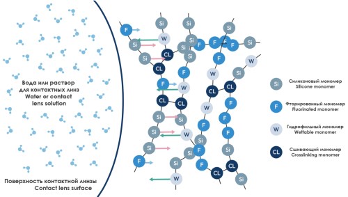
This article explores how modern contact lens materials have evolved from heavy, oxygenblocking early plastics to advanced polymers that allow high oxygen flow, better comfort, and improved durability. Key innovations such as hydrogels, silicone components, and fluorinated monomers have greatly enhanced wettability, stability, and overall eye health. These developments continue to support safer and more effective lenses, including specialised designs like scleral and orthokeratology lenses.

Modern contact lenses (CLs) provide a convenient and comfortable alternative to spectacles. Prolonged CL wear leads to the formation of protein–lipid deposits, the composition of which depends on lens material properties and tear film composition. The removal of protein deposits and bacterial contamination is a primary function of multipurpose solutions (MPS), ensuring safe contact lens wear and helping to prevent microbial colonization. The proposed method for visualizing protein deposits enables ophthalmologists to assess the degree of contact lens surface contamination and to evaluate the cleaning efficacy of multipurpose solutions.
DISCUSSION CLUB
NEWS: WHAT? WHERE? WHEN?