


ORIGINAL ARTICLES
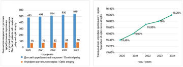
Introduction. Optic nerve atrophy (ONA) holds a leading position in the nosological structure of ophthalmopathology. In children with cerebral palsy (CP), the presence of concomitant pathology such as ONA significantly worsens their health and quality of life. The combination of ONA and CP, as well as the interrelationship of these diseases, represents a relevant subject for research, particulary in studying the clinical and epidemiological features of ONA in children with CP to find optimal clinical solutions and prognostic approaches. Purpose: to investigate the prevalence of optic nerve atrophy in children with cerebral palsy to determine the prognosis for the development of this pathology in the study group. Materials and methods. A retrospective analysis of medical records of 640 patients diagnosed with cerebral palsy who were hospitalized in the clinic from 2015 to 2020 was conducted. The analysis included data from children with early-onset cerebral palsy (up to 3 years old) who were hospitalized for the first time. MS EXCEL software was used to calculate the main indicators of linear regression. Results. The study revealed that optic nerve atrophy in young children develops more frequently in boys (1.5 times more often than in girls), in rural areas, and in those with spastic cerebral palsy (36.3 %). The second most common form was ataxic cerebral palsy (69 children, 21.6 %), followed by infantile hemiplegia (61 children, 19%). Based on the dynamics of the prevalence of optic nerve atrophy in children with cerebral palsy, a mid-term forecast was made using the main indicators of linear regression. The prevalence of optic nerve atrophy in children with cerebral palsy is increasing, with prognostic estimates indicating further growth (14.9% in 2015, 15.4% in 2020, and 16.2% in 2024). Conclusion. The results of the study showed that cerebral palsy in children is often associated with optic nerve atrophy. ONA in young children is more frequently developed in cases of spastic cerebral palsy. As a result of calculating, an increase in the prevalence of optic nerve atrophy in children with cerebral palsy was observed over time, based on the correlation calculated using Spearman’s non-parametric correlation analysis Prognostic estimates indicate a significant increase in the near future.
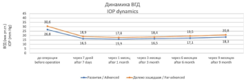
Introduction. Glaucoma is one of the most common visual pathologies leading to blindness and low vision worldwide. Refractory glaucoma is characterized by a severe course and resistance to traditional treatment methods. One of the promising treatments for such patients is micropulse transscleral cyclophotocoagulation (mCPC), which allows laser energy to be delivered to a localized area in doses without causing thermal damage to the structures of the ciliary body. With the advent of this method, new opportunities have emerged in the treatment of patients not only in the terminal stages of glaucoma with residual visual functions but also in the earlier stages of the disease with preserved and even high vision. Purpose: to evaluate the effectiveness of mCPC in patients with refractory, operated glaucoma with preserved visual functions using ultrasound biomicroscopy (UBM). Materials and methods. From April 2023 to January 2024, 80 patients aged 68,3 ± 5,6 years with refractory, operated open-angle glaucoma were examined and prescribed mCPC using the Quantel Medical device (Supra 810, France). The patients were divided into 2 groups: Group I consisted of 32 patients (32 eyes) with advanced glaucoma, while Group II included 48 patients (48 eyes) with far-advanced glaucoma. All patients were on the maximum antihypertensive regimen and had a history of repeated laser and surgical interventions. The procedure was carried out using standardized technology. Results. In the early postoperative period, a hypothensive effect was achieved in all cases, with an average intraocular pressure (IOP) decreased by 38%. The antihypertensive regimen was discontinued in 37% of patients. By the 9th month, 55% of patients had reduced the number of medications used, and 45% of patients in the second group underwent repeated mCPC, after which the IOP decreased to 16,7 ± 0,06 mm Hg in this group. Conclusion. mCPC is an effective and safe method for treating refractory, operated glaucoma, leading to a persistent decrease in IOP without affecting visual functions and the structure of ciliary body. It also reduces the number of used antihyperthensive drugs used.
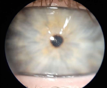
Introduction. Keratoconus is a progressive corneal disease that results in thinning, curvature change, and refractive alterations, significantly impairing. visual acuity, even to the point of light perception. In advance stages, corneal transplantation may be necessary. Purpose. This publication aims to analyze a clinical case of corneal transplantation in a patient with keratoconus and Stormorken Syndrome, a rare genetic disorder caused by mutations in the STIM1 gene leading to calcium channelopathies. Case description. Patient Z., a 24-year-old male diagnosed with Stormorken Syndrome in childhood, was referred to the Moscow City Ophthalmological Centre. Comprehensive evaluations, including biomicroscopy, keratopachimetry, and ultrasound eye examination, confirmed keratoconus in both eyes. The patient underwent bilaterial penetrating keratoplasty due to disease progression. Postoperatively, visual acuity was 0.8 in the right eye, and 0.7 in the left eye. Additional clinical observations including bilateral myositis (resistant to medication), hypoinflammation, thrombocytopenia, dyslexia, and proximal muscles weakness, of the consistent with Stormorken Syndrome. Genetical testing confirmed a pathogenic STMI1 mutation. Conclusion. Despite the high volume of keratoplasty procedures at our clinic, this case is noteworthy. The currrent literature lacks reports on penetrating keratoplasty using native donor material in patients with Stormorken Syndrome, highlighting the uniqueness of this case.
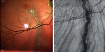
Introduction. Choroidal macrovessel (CM) is a rare anatomical feature most often detected incidentally. Recent studies suggest an arterial origin of CM, with the majority of authors considering it to be an enlarged short posterior ciliary artery. The literature describes both variants of CM not associated with retinal changes and those complicated by exudative manifestations. Purpose. This study aims to describe two clinical cases of choroidal macrovessel and conduct a differential diagnosis based on literature data with clinically similar conditions. Material and methods. Patients underwent a standard ophthalmologic examination, as well as optical coherence tomography, optical coherence tomography angiography (protocols 10 and 20 degrees), and multispectral laser scanning. Results. We present descriptions of two clinical cases: CM in a patient with choroidal folds and a prominent CM with accompanying subretinal fluid. In both cases, CMs were incidental findings not associated with reduced visual acuity. In the second clinical example, we demonstrate the presence of subretinal fluid in a patient with CM, with no negative progression after one year of observation. For differential diagnosis, an analysis of conditions with similar clinical manifestations was conducted based on literature data. Conclusion. The presented clinical cases illustrate the diversity of CM manifestations. In most cases, the pathology is not associated with retinal structural abnormalities; however, it can cause retinal changes in some patients. Due to its rarity, CM can be considered a poorly studied anatomical feature, warranting dynamic observation upon detection.
REVIEWS
Introduction. An increasing number of patients with a history of keratorefractive surgeries are presenting to ophthalmologists with complaints of vision loss due to cataracts. Treating this group poses surgeons with a range of unique challenges: high demands for vision quality, complexities in selecting the appropriate intraocular lens (IOL) power calculation formula and IOL model, target refraction, as well as the need to modify cataract extraction techniques and address specific postoperative considerations. Despite advancements in the development of new IOL designs and calculation formulas, clinical and functional outcomes in this group remain inferior to those in patients without prior keratorefractive procedures. A paradigm shift is emerging, advocating for a personalized approach in the diagnosis and management of cataracts in these patients. However, discussing all aspects within a single review proved impractical, leading us to divide it into two parts. The objective of the first part of this study is to assess the specific considerations for aphakia correction in patients who have undergone keratorefractive procedures, based on literature data, while taking into account the long-term complications of refractive surgery. Additionally, this part will address the fundamental principles of the design and functionality of pseudoaccommodating intraocular lenses (IOLs). Materials and methods. A selection of over 200 peer-reviewed publications from resources such as PubMed, eLibrary, CyberLeninka, Science Direct, and Google Scholar over the past 30 years was conducted. The first part of the review includes 49 publications. This work represents an analysis of contemporary literature, reflecting the impact of keratorefractive surgeries on the successful performance of phacoemulsification with IOL implantation. Results. The findings from the first part of the analysis indicate that a detailed medical history of previously performed keratorefractive corrections – specifically their type and potential long-term complications – play a significant role in determining the surgical treatment strategy. Standard examination methods do not always fully reflect the optical characteristics of the cornea in these patients. Extended preoperative assessments, including specialized techniques such as keratotopography and keratotomography, are crucial for identifying corneal irregularities and for the subsequent selection of the type of intraocular lens (IOL) for aphakia correction in patients who have undergone keratorefractive surgeries. Studies show high effectiveness not only in using monofocal lenses but also in the potential application of pseudoaccommodating IOLs, including those with extended depth of focus and multifocal lenses. The selection of optimal formulas for IOL calculation, as well as the clinical aspects influencing refraction in the postoperative period, will be addressed in the second part of the literature review. Conclusion. The increase in the number of refractive surgeries has led to a growing population of patients with cataracts following ametropia correction. This has spurred the development of new IOL variants with extended depth of focus. However, literature data on their effectiveness in patients who have undergone keratorefractive procedures remain limited. Multicenter prospective studies are needed to evaluate new IOL models and to determine the optimal surgical strategies for this category of patients.
Introduction. The term “Dysfunctional Lens Syndrome” (DLS) refers to age-related changes in the lens, including early cataracts and presbyopia, that do not significantly reduce visual acuity. This syndrome is characterized by a decrease in accommodative amplitude, an increase in light scattering, and a reduction in contrast sensitivity. With rising life expectancy, the prevalence of DLS is increasing, necessitating a more in-depth study of the pathogenic mechanisms underlying the syndrome and the refinement of its diagnostic criteria to establish standards for treatment correction. Aim: to summarize information on the pathogenesis of DLS, the potential for staging the syndrome using objective examination methods, and to provide treatment recommendations. Materials and methods. A bibliographic study of scientific publications was conducted using the Pubmed, ScienceDirect, and Cyberleninka databases. Literature sources were searched using the following keywords: dysfunctional lens syndrome (DLS), presbyopia correction, cataract, age-related lens changes. A total of 32 publications, primarily from the last 10 years, were included in the study from more than 100 viewed sources. Results. The DLS encompasses a wide range of conditions, from early stages characterized by a loss of accommodative amplitude and minor refractive anomalies to later stages involving a decrease in visual acuity and quality due to increased light scattering and aberrations. Currently, the evaluation of age-related lens changes involves assessing visual acuity and determining the degree of lens opacification using slit-lamp examination. Additionally, objective instrumental methods, such as optical coherence tomography and Scheimpflug imaging, can be used to assess the degree of lens opacification. Various methods are employed for the correction and treatment of DLS, ranging from glasses and contact lenses to surgical interventions, such as lens extraction and intraocular lens implantation. Research is also ongoing into drugs aimed at slowing the progression of lens changes. Conclusion. The term “Dysfunctional Lens Syndrome” is currently used to describe early cataracts and presbyopia, where the reduction in visual acuity is still minor, but patients experience visual complaints due to age-related lens changes. Further research is necessary to develop and standardize diagnostic criteria and to evaluate the effectiveness of new treatment methods and interventions in order to create the most effective and adequate approach to correcting DLS and alleviating the associated visual complaints.
TECHNOLOGIES

In this article, we will examine the importance of oxygen permeability (Dk) in gas permeable contact lens materials, and the significance of high Dk in scleral and ortho-k lens designs.
WORKSHOP
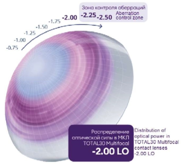
Multifocal soft contact lenses are the fastest growth segment in contact lenses. While presbyopic patients demonstrate an increasing interest in contact lenses, not all practitioners actively integrate multifocal lenses into their practices. Some of the reasons include the perception that multifocal fitting is time-consuming and chances of successful adaptation of “ageing” patients are low. The purpose of this publication is to provide an overview of the latest multifocal contact lens, essential fitting steps and instruments for quick and effective multifocal lens fitting.


