


ANNIVERSARIES

ORIGINAL ARTICLES
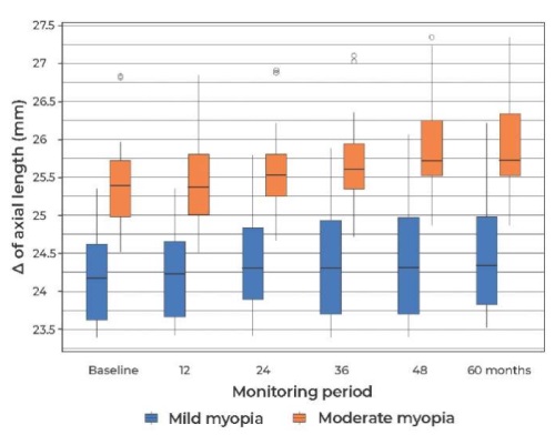
Introduction. Myopia is a widespread and growing public health concern. The impact of soft contact lenses (MCLs), which create a relative myopic peripheral defocus with an add power of +1.5 to +2.00 D, on slowing myopia progression has been widely studied in short- and long-term studies. However, studies lasting more than three years that evaluate the effect of high add multifocal soft contact lenses (MFCLs) on ocular growth and refractive error dynamics are limited. Purpose: to evaluate the effect of bifocal soft contact lenses (BFCLs) Prima BIO Bi-focal with an add power of +4.00 D on the dynamics of refractive error and axial eye length in patients with progressive myopia over a prolonged period of wear. Materials and methods. Twenty-eight patients (28 eyes) with bilateral myopia, with a spherical equivalent (SE) refractive error between –0.75 and –5.5 D, astigmatism <1.25 D, and anisometropia up to 1.00 D, were included in this study conducted from 2018 to 2023. The mean age of the participants was 10 years. Based on the degree of myopia, the patients were divided into two groups: the first group consisted of 13 patients with mild myopia (–0.75 to –3.00 D), while the second group consisted of 15 patients with moderate myopia (–3.25 to –5.50 D). Cycloplegic refraction, visual acuity, and axial eye length were evaluated at the initial examination and follow-up visits at 12, 24, 36, 48, and 60 months. Patients in both groups wore BFCLs Prima BIO Bi-focal with an add power of +4.00 D on a monthly regimen for at least 10 hours per day. Results. After 60 months of wearing high add BFCLs, the change in refractive error from baseline was 0.25 (0; 0.75) D in the first group and 1.25 (0.5; 1.5) D in the second group. The increase in axial eye length after 60 months compared to baseline was 0.14 (0.03; 0.24) mm in the group with mild myopia, and 0.48 (0.21; 0.55) mm in the group with moderate myopia. Conclusion. The data indicate a stabilizing effect of high add BFCLs on the progression of both mild and moderate myopia. A greater antimyopic effect, in terms of refractive error dynamics and axial elongation, was observed in the group with mild myopia. Further research is needed to explore the correlation between the antimyopic effect of BFCLs and the degree of myopia.

This article evaluates the clinical benefits of using a high Dk material such as Optimum Infinite (180 Dk) material in orthokeratology, demonstrating faster myopia correction due to its higher oxygen permeability, which reduces corneal edema.
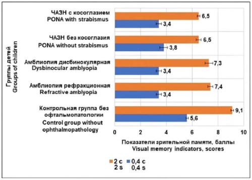
Introduction. Visual impairments present significant challenges for learning in school, limit career choice, and restrict participation in sports. Therefore, the developing a comprehensive diagnostic system for assessing visual functions in children with functional and organic ophthalmopathology is a pressing tasks in modern ophthalmology. The aim is to further advance effective treatments and medical-pedagogical support for such children. Purpose: to conduct a comparative analysis of visual function indicators in schoolchildren with partial optic nerve atrophy (PONA) and amblyopia in order to create an effective diagnostic system for detecting monocular and binocular visual function disorders. Materials and methods. The study observed 120 school-age children with PONA, of whom 85 had strabismus and 35 did not. The group of schoolchildren with amblyopia consisted of 98 children (40 with dysbinocular amblyopia and 58 with refractive amblyopia). The control group included 67 children without ophthalmopathology. In addition to standard ophthalmological examinations, electrophysiological parameters (electrical sensitivity threshold and electrical lability), fusion amplitude, binocular and stereovision presence, stereokinetic effect, and visual memory indicators were assessed. Results. The study revealed that, along with organic causes of visual impairment (such as significant decreases in visual acuity, narrowing of visual fields, and worsening electrophysiological parameters), children with PONA also exhibited binocular visual function disorders. These included reduced fusion amplitude, absence of binocular and stereovision, and dominance of monocular spatial perception mechanisms. Binocular disorders were observed in children both with and without strabismus, while among children with amblyopia, these disorders are mainly associated with the dysbinocular form. A decrease in visual memory indicators was identified in both children with PONA and those with amblyopia. Conclusion. Binocular vision disorders and reduced visual memory were observed in schoolchildren with both partial optic nerve atrophy and amblyopia. However, binocular visual function disorders in children with PONA were identified both in the presence of strabismus and orthotropy, unlike children with amblyopia, who primarily exhibited binocular disorders in the presence of strabismus. The inclusion of visual memory and spatial perception indicators in the diagnosis and monitoring of visual functions supports a more individualized approach to the medical and pedagogical care of children with ophthalmopathology.
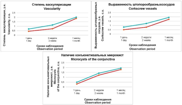
Introduction. One limitation of glaucoma surgery is its reduced long-term effectiveness due to scarring of intraocular fluid outflow pathways. Appropriate perioperative therapy may extend the hypotensive effect by reducing scarring and improving the filtration bleb. Purpose: to evaluate the effect of perioperative use of Oku-Oku® on intraocular pressure (IOP) and filtration bleb condition in the early postoperative period following glaucoma surgery. Materials and methods. Thirty patients (30 eyes) who underwent non-penetrating glaucoma surgery (NPGS) were studied. The main group (15 eyes) received Oku-Oku® drops twice daily: the day before surgery, immediately before surgery, and for five days postoperatively. The control group (15 eyes) received no additional medication. Tonometry and assessment of conjunctival hyperemia were performed the day before surgery, and at 1 day, 2 weeks, and 1 month postoperatively. Filtration bleb characteristics were assessed using the Würzburg Clinical and Morphological Classification of Filtration Pads at the same time points, except for the preoperative assessment. Results. Glaucoma surgery significantly reduced IOP in both groups. After 1 month, the mean IOP in the main group was lower (15.1 ± 4.1 mm Hg) compared to the control group (17.9 ± 3.3 mm Hg). Conjunctival hyperemia was also lower in the main group (15.4 ± 2.7%) than in the control group (18.0 ± 3.1%). Filtration bleb parameters improved in both groups over time, with the greatest differences observed at 1 day and 2 weeks post-surgery. Conclusion. Perioperative use of Oku-Oku® reduced conjunctival hyperemia in the filtration bleb area during the first month after glaucoma surgery. This early improvement in the intraocular fluid deposition area may enhance the effectiveness of glaucoma surgery. Oku-Oku® can be used in comprehensive preparation for glaucoma surgery and other similar anterior segment procedures.
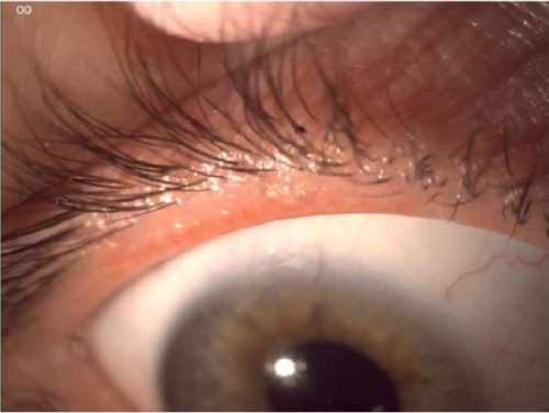
Introduction. Acne is a chronic, recurrent, and inflammatory skin disease that that can have significant aesthetic and psychological impacts on patients. The first-line treatment for acne is isotretinoin, which exerts systemic effect, reducing sebum production and alleviating inflammation However, one of the most common complications associated with prolonged systemic retinoid use is dry eye syndrome, linked to meibomian gland dysfunction, warranting special attention from ophthalmologists. Purpose: to present a clinical case of a patient who underwent four months of systemic isotretinoin therapy for acne and subsequently developed ocular complaints. Methods. Comprehensive ophthalmological diagnostics were conducted, including patient history, biomicroscopy, and corneal staining with fluorescein. Additional assessment included non-invasive tear film breakup time, measurement of tear meniscus height, evaluation of the lipid layer thickness of the tear film, meibography, eyelid margin photography, conjunctival hyperemia analysis, and corneal fluorescein staining. Treatment included various groups drug classes such as moisturizing drops, reparative gels, corneal protectors, and glucocorticosteroids. A therapeutic eyelid massage was also performed. Results. After three months of active therapy, the patient exhibited significant improvement, with a marked reduction in subjective complaints, no corneal epithelial defects, and an increase in tear film breakup time. Long-term symptom remission was achieved after five months of therapy. Conclusion. This case highlights the importance of interdisciplinary collaboration between ophthalmologists and dermatologists in managing patients undergoing long-term systemic retinoid therapy for acne. Comprehensive ophthal- mological assessment using specialized diagnostic methods is essential for accurately evaluating the clinical picture of ocular conditions and administering timely, multi-faceted therapy. This approach enhances patients’ quality of life and helps prevent severe complications.
REVIEWS
Introduction. In clinical practice, cataract surgeons are increasingly encountering patients with a history of keratorefractive surgery. According to various studies, one of the key challenges in achieving the desired refractive outcome for these patients is selecting the appropriate formula for intraocular lens (IOL) power calculation. The second part of this review explores the application of different formulas for calculating IOL power, with a focus on specific challenges and potential errors in IOL selection for aphakia correction following previous keratorefractive procedures. The purpose of this second part is to address the issue related to IOL selection and calculation in patients post-keratorefractive surgery, considering clinically significant optical effects and the condition of the anterior surface of the eye, which can influence both postoperative refraction and the duration of the postoperative rehabilitation period. Materials and methods. A review of peer-reviewed publications from the past 30 years was conducted using databases such as PubMed, eLibrary, CyberLeninka, Science Direct, and Google Scholar. The review analyzed 32 articles, predominantly from the last decade. This study provides a detailed analysis of the scientific literature, highlighting the influence of prior keratorefractive surgeries on the success of phacoemulsification with IOL implantation. Results of this second part of the review suggest that using multiple IOL calculation formulas yields more accurate refractive outcomes, minimizing postoperative refractive errors. Additionally, the analysis of IOL selection criteria highlights that IOLs with extended depth of focus (EDOF) demonstrate greater tolerance to decentration, pupil size variations, and higher-order aberrations, facilitating patients adaptation to improved visual quality following cataract surgery. Early diagnosis and effective treatment of underlying dry eye syndrome (DES) can further reduce errors during preoperative diagnostics and enhance patient satisfaction in the post-operative period. Conclusion. Taken together, both parts of this article provide a comprehensive review that, for the first time, brings together key fundamental and clinical aspects of aphakia correction in patients with a history of keratorefractive surgery. Understanding these factors will assist ophthalmic surgeons in selecting optimal treatment strategies to achieve the best possible clinical outcomes for these patients.
WORKSHOP
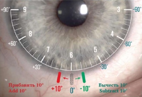
Uncorrected astigmatism is one of the most common causes of reduced visual acuity in the world population. The optical system of the eye, in which the presence of clinically significant astigmatism (more than 1.0 D) is detected, is quite common. In the absence of adequate correction or inadequate diagnosis, astigmatism significantly reduces the uncorrected visual acuity of patients, which affects their quality of life. Eyeglass and contact vision correction are the most common methods of non-surgical correction of ametropia. However, according to the data of foreign and Russian studies of the contact astigmatism correction market, the share of toric lenses fitting is only a small part among all prescriptions.
This article is devoted to detailing of the fitting algorithm of new toric soft contact lenses manufactured by OKVision® Fusion NEW Toric and aims to help optometrists to fit this type of lenses more confidently, which in turn will help users to obtain full- fledged vision correction.


