ORIGINAL ARTICLES
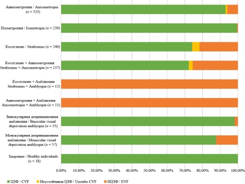
Introduction. The prevalence of amblyopia is reported to reach 6% among children and 5.6% among adults. Patients with amblyopia exhibit reduced reading speed, impaired learning abilities, disrupted fine motor skills, and diminished or absent stereopsis. The state of visual fixation significantly impacts treatment outcomes, as children with central visual fixation (CVF) and eccentric visual fixation (EVF) require different management strategies. However, there is a lack of analytical data regarding the prevalence of EVF among patients with various ophthalmic pathologies and methods for its evaluation. Objective: to identify and recommend the most common method for assessing visual fixation for clinical practice and to determine the prevalence of EVF among children with different ophthalmic condition. Materials and methods. This study reviewed the findings of both Russian and international research on the prevalence of EVF in children. The review included 30 experimental, observational, retrospective, and comparative publications that provided data on visual fixation status in patients with various ophthalmic conditions and the methods used to assess it. Results. Analysis of full-text articles led to the selection of eight studies published between 2017 and 2024 and one from 2009. These studies encompassed data on the ophthalmic status, visual fixation, and methods of assessment for 1,510 individuals. Among patients with anisometropia, 93.1% exhibited CVF, 6% had EVF, and five patients demonstrated unstable CVF. For patients diagnosed with strabismus without concomitant pathology, 74.2% had CVF, 21.7% had EVF, and 4.2% showed unstable CVF. Among patients with strabismus combined with anisometropia, CVF was found in 72.3%, EVF in 25.5%, and unstable CVF in 2.2%. The highest prevalence of EVF (27.5%) was observed among children with amblyopia and/or strabismus. The most commonly utilized and accessible method for assessing visual fixation across nine studies was direct ophthalmoscopy, performed in three-quarters of the cases. Conclusion. EVF is most frequently encountered in children with strabismus and amblyopia. The use of direct ophthalmoscopy as a diagnostic method for EVF is accessible for any ophthalmology office and is recommended for widespread implementation in clinical practice.
The exudative form of age-related macular degeneration (AMD) requires differential diagnosis from centrally localized choroidal neoplasms, particularly choroidal melanoma. It is important to consider that these conditions can often coexist, complicating the diagnostic process. Studies dedicated to the use of optical coherence tomography (OCT) for differentiating AMD and choroidal tumors are scarce. Objective: to determine the structural OCT differences between exudative AMD, choroidal melanoma (CM), and choroidal hemangioma (CH) localized in the central fundus area. Materials and methods. The study included 60 patients referred for consultation with suspected centrally localized choroidal neoplasms. The average age at the time of examination was 60.75 ± 15.36 years; the mean prominence of the lesion according to ultrasound (US) was 1.81 ± 1.44 mm, and the average diameter was 8.31 ± 1.44 mm. All patients underwent OCT in standard and Enhanced Depth Imaging (EDI) modes using the OCT Spectralis (Heidelberg Engineering, Germany). OCT scans were analyzed for the condition of the “choroidal complex”, the choroid in the area adjacent to the lesion, the integrity of Bruch’s membrane, the state of the retinal pigment epithelium (RPE), the presence or absence of neuroepithelial detachment (NED) and cystic changes, as well as the ability to differentiate retinal layers. Results. For AMD, the key differential diagnostic OCT criterion is the absence of choroidal elevation, with significant retinal changes at the surface level. These changes are characterized by RPE detachment, extensive hyperreflective foci (ophthalmoscopically corresponding to scarring), and destructive changes in the overlying retinal layers. OCT features of choroidal tumors include elevation of the “choroidal complex” with less pronounced changes in the overlying retina. The structure of the “choroidal complex” varies depending on the nature of the pathological process. In CM and CH, changes in the RPE and the adjacent retina are less pronounced and occur secondarily due to choroidal vessel compression by the growing tumor and their obliteration in CM or pathological changes in the intrinsic choroidal vessels in CH. Conclusion. In the differential diagnosis of AMD and centrally localized choroidal neoplasms, attention should be paid to the condition of the “choroidal complex” and Bruch’s membrane in the area of interest when analyzing OCT findings.
Introduction. The gold standard for surgical treatment of patients with advanced proliferative diabetic retinopathy (PDR) is vitreoretinal surgery (VRS). However, the optimal timing for the removal of early cataracts in this patient category remains an open question. Objective: to evaluate the effectiveness of phacoemulsification of complicated early cataracts as a second stage after VRS in patients with advanced proliferative diabetic retinopathy PDR. Material and methods. The study included 216 patients diagnosed with PDR and complicated early cataracts. These patients were divided into four groups based on the surgical approach. Group I (n = 77): Stage 1 – vitreoretinal surgery (VRS) with silicone tamponade: Stage 2 – phacoemulsification of cataracts (PhEC) with intraocular lens (IOL; implantation performed simultaneously with silicone oil removal. Group II (n = 76): Stage 1 – PhEC with IOL implantation combined with VRS and silicone tamponade; Stage 2 – silicone oil removal from the vitreous cavity. Groups I and II were further divided into two subgroups. Subgroup Ia (n = 62): operations were performed as described above. Tear fluid samples were collected from some patients (n = 17) before surgery and on the second day after Stage 1. Subgroup Ib (n = 15): patients received an intravitreal injection of an angiogenesis inhibitor 10–14 days before VRS in a single dose of 0.5 mg. Subgroup IIa (n = 62): operations were performed as described above. Tear fluid samples were collected from some patients (n = 17) before surgery and on the second day after Stage 1. Subgroup IIb (n = 14): patients received an intravitreal injection of an angiogenesis inhibitor 10–14 days before VRS in a single dose of 0.5 mg. Group III (n = 32): Stage 1 – VRS with gas-air tamponade. Stage 2 – PhEC with IOL implantation. Group IV (n = 31): PhEC was performed simultaneously with VRS using a gas-air mixture for tamponade. Results. Patients in subgroup Ia and Group III showed better visual function outcomes compared to those in subgroup IIa and Group IV, respectively (p < 0.001). The intensity of the inflammatory response (2–3 points) was significantly higher in subgroup IIa (p < 0.001) and Group IV (p < 0.001) compared to subgroup Ia and Group III, respectively. Neovascular glaucoma was significantly more common in subgroup IIa patients (n = 9; 14.5%) compared to subgroup Ia (n = 2; 3.2%, p = 0.027). Similarly, the incidence of neovascular glaucoma was higher in Group IV (n = 6; 19.3%) compared to Group III (n = 1; 3.1%, p = 0.04). Subgroup IIa showed 2–2.5 times higher concentrations of IL-8, MCP-1, and ICAM-1 molecules compared to subgroup Ia. Conclusion. Phacoemulsification of early cataracts as a second stage after VRS in patients with advanced PDR provides a gentle approach to surgical treatment for this category of patients. This approach improves anatomical and functional outcomes and reduces the number and severity of postoperative complications.
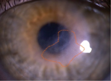
Relevance. The use of scleral contact lenses (SCLs) is currently indicated for patients with complex ametropia and irregular corneal surfaces resulting from various causes. SCLs are effective in cases of intolerance to other vision correction methods or when glasses or standard contact lenses fail to provide satisfactory visual function and optical rehabilitation. A critical factor in SCL design is ensuring sufficient oxygen transmission for safe wear, which can be enhanced by fenestration. The study aims to demonstrate the possibility of optical rehabilitation in a post-keratitis patient using fenestrated scleral lenses made of Optimum Infinite material. Objective: to demonstrate the possibility of optical rehabilitation in a post-keratitis patient using fenestrated scleral lenses made of Optimum Infinite material. Materials and methods. A patient born in 2013 presented with complaints of low visual acuity. The patient’s history included keratitis, leading to ametropia uncorrectable with glasses. In addition to standard examinations, corneal topography, pachymetry, and morphological evaluation via scanning confocal microscopy were performed. Results. A scleral contact lens (OKVision SMARTFIT®) made of high-oxygen-permeable Contamac Optimum Infinite material with fenestration (1 × 0.3 mm) was fitted. The patient’s visual acuity improved from 0.08 to 0.9 after lens fitting. No complications associated with lens wear were observed during the follow-up period. Conclusions. Scleral contact lenses made from the high-oxygen-permeable material Contamac Optimum Infinite with fenestration can be recommended for optical correction in patients with ametropia induced by keratitis.
REVIEWS
Introduction. Memory is at the core of all intellectual activities; no cognitive function can be performed without its involvement. However, the mechanisms underlying its function remain incompletely understood. Objective: to review publications on current perspectives regarding the mechanisms of human visual memory and identify promising directions for future research in this area. Materials and methods. A literature analysis was conducted based on 58 publications from the last 15 years, sourced from Google Scholar, PubMed, eLibrary, and Crossref Metadata Search. Results. This review highlights the distinctions between longterm and working visual memory based on their functional properties, which rely on different neural substrates. Working memory mechanisms are associated with the activity of the occipital and parietal cortices, while long-term memory is linked to the medial temporal lobe and hippocampus. Traditionally, the organization of long-term visual memory has been modeled as a passive storage system, retaining visual information for extended periods. However, contemporary models of working visual memory describe it as a cognitive mechanism for retrieving necessary information from long-term memory and applying it to functional tasks. Both models emphasize the interaction of hypercolumn neurons, various layers of the visual cortex, and cerebellar structures in evaluating color, spatial localization, and other visual characteristics. Conclusion. Despite extensive and multifaceted research in recent years, many aspects of visual memory remain insufficiently explored. One promising direction for future studies is investigating the functioning of visual memory across different age groups in patients with ophthalmic pathology, including conditions affecting the central visual processing system.

Scleral contact lenses are considered one of the most effective methods of optical correction for induced ametropia, particularly in cases of irregular corneal surface. Advancements in scleral lens materials and design have significantly expanded the indications for their fitting. However, ophthalmologists have insufficient awareness of this correction method and its potential in the optical rehabilitation of patients. Objective: to review and summarize the literature on the historical milestones in the development of scleral contact lenses and their application in modern ophthalmic practice. Methods. A literature analysis was conducted using the eLibrary and PubMed databases, covering publications from the last 30 years. Over 200 relevant articles were reviewed, of which 50 were included in this study. Results. This review outlines the history of scleral lens development, detailing key stages in their evolution. The characteristics of modern scleral lenses, including the materials used and the equipment for their fabrication, are described. The indications and contraindications for scleral lens fitting are also discussed. Conclusion. Scleral lenses are now widely available and are custom-manufactured based on individual patient parameters using highly gas-permeable materials. Their use enables optimal visual outcomes even in challenging clinical cases where other correction methods prove ineffective.
Relevance. Myopia is a significant issue in ophthalmology and public health. Its progression not only leads to a decline in visual function and reduced quality of life but also increases the risk of vision-threatening complications. In recent years, alongside contact lens-based approaches that slow excessive eye growth, pharmacological, optical, and behavioral myopia control strategies have been actively developed. Objective: to summarize and systematize data from randomized clinical trials conducted over the past five years on the efficacy of non-invasive myopia control methods, identifying the most effective approaches and their combinations. Materials and methods. A literature search was conducted in the eLibrary, PubMed, and Scopus databases using the keywords “Myopia Control” and “progressive myopia”. A total of 3,714 studies published between 2020 and 2025 were identified, of which 52 full-text articles met the inclusion criteria. The final analysis included 36 publications that provided data on both refractive error progression and axial length changes. Results. The use of defocus-incorporated multiple segment (DIMS) and other myopia control spectacle lenses has demonstrated high efficacy in slowing axial elongation, confirming the clinical significance and promise of this approach. However, questions remain regarding the magnitude and consistency of their long-term efficacy. Low-dose atropine has shown the greatest effect when combined with single-vision spectacle correction. However, the variability in results due to different atropine concentrations necessitates further research. Increased time spent outdoors has been associated with a reduced risk of myopia progression in individuals with pre-myopia and low myopia. The combination of red-light therapy with spectacle correction has proven more effective than red-light therapy alone. While these methods show promising results, long-term studies are required to confirm their efficacy and safety. Conclusion. Effective myopia control in children requires the development of combined strategies incorporating optical, pharmacological, and behavioral interventions. Despite the availability of effective non-invasive myopia control methods, questions remain regarding their mechanisms of action and the long-term efficacy of combination treatments.
TECHNOLOGIES
Optometry, as a branch of ophthalmology, has expanded significantly in Russia over the past three decades. Although the training of optometrists dates back to the Soviet era, the educational program then primarily focused on producing corrective devices rather than fitting them, and optometrists had little involvement in diagnostic or therapeutic processes. Demographic changes linked to an aging population, along with rising rates of refractive disorders, have significantly increased the workload for ophthalmology services throughout Russia. Another critical challenge in managing ocular pathologies is the shortage of ophthalmologists. One effective strategy to address these issues is to organize a more active role for optometrists in the healthcare system, yet this requires high-quality specialist training within secondary vocational education (SVE). Despite the updated educational standard for the specialty of medical optician-optometrist, there are still no well-established teaching methods for optometry in the SVE system, and no textbook has been approved by the Ministry of Education of the Russian Federation. Consequently, selecting and adapting contemporary teaching technologies within the educational project design for preparing qualified optometrists is a vital task in the SVE program. Educational technologies serve as a mechanism to orginize innovative forms and methods of instruction, bringing them to life in authentic pedagogical practice. Recognizing project design as an effective tool for modern education highlights the need to integrate independent and creative student activities into the main educational process. This article explores various approaches to designing the educational process for medical opticians-optometrists in the SVE program.

The past 15 years have witnessed a remarkable increase in the use of scleral lenses worldwide, with some regions experiencing more rapid adoption than others. This article examines the factors contributing to this growth and considers whether similar advancements can be achieved in regions with slower initial progress. Advancements in materials, improvements in diagnostic technology, and systemic market influences have all played a role in shaping the global scleral lens market.
WORKSHOP
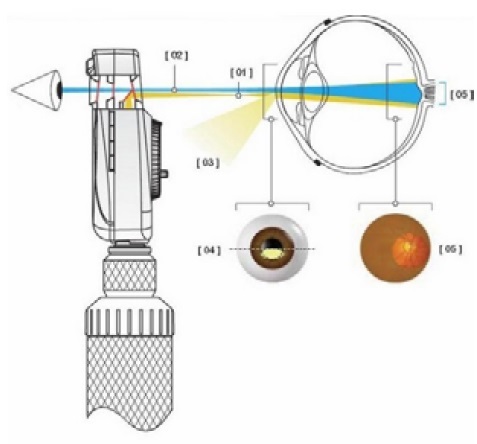
Assessment of visual fixation parameters is an essential diagnostic method for patients with amblyopia. Determining the type of visual fixation helps predict the clinical outcome of amblyopia and serves as the foundation for developing treatment strategies and tactics. This article aims to familiarize ophthalmologists with an algorithm for determining visual fixation in a simple, objective, and rapid manner. The practical section covers the diversity of retinal reflexes, introduces the concept of visual fixation, and reviews both national and international classifications. Special attention is given to the algorithm for determining visual fixation using a direct ophthalmoscope and the fundamental principles for interpreting the obtained results.