ORIGINAL ARTICLES
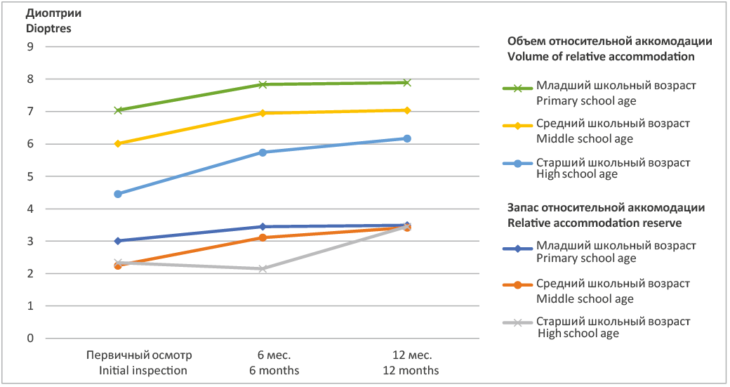
Relevance. Myopia is one of the most common refractive pathologies in the world. By 2050, according to WHO 2015, more than half of the world’s population will suffer from myopia, which will lead to the risk of developing pathologies associated with the progression of myopia, such as glaucoma, macular degeneration and retinal detachment. The use of various modern methods to control myopia can slow down the progression of myopia, which potentially reduces the risks of complications. Objective: to evaluate changes in the anatomical and functional parameters of the eye (clinical refraction, axial length, accommodation functions) in children with progressive myopia corrected with glasses with Stellest lenses in different age groups. Materials and methods. The study was conducted on the basis of the Eye Microsurgery Clinic of the Stavrapol State Medical University. Glasses with Stellest lenses were assigned to 80 children aged 8 to 16 years. The average age of the children was 11 ± 0.12 years. The children were divided into three age groups: 1st – primary school; 2nd -middle school; Group 3 – senior school. After the examination, the children were selected glasses with Stellest lenses. The average period of wearing glasses with Stellest lenses was 12 months. Clinical refraction, axial length and accommodation functions were evaluated during the observation. Results. The change in refraction depended on the age of the child. The greatest increase in refraction by 0.40 ± 0.02 D was observed in the younger age group, and the smallest (0.27 ± 0.02 D) in children of secondary school age. In the group of children of senior school age, the increase in refraction was +0.32 ± 0.03 D. The axial length of the eye in children of the primary school group after 12 months of wearing glasses with Stellest lenses signifi cantly increased by an average of 0.28 ± 0.03 mm. This axial growth of the eye correlates with an increase in myopic refraction in the same group of children. In the group of children of senior school age, the growth of PZO was 0.1 ± 0.04 mm. There was an increase in the accommodation reserve, the positive relative accommodation and the relative accommodation in all groups. Conclusions. The study showed that wearing glasses with Stellest lenses helps to reduce the rate of progression of myopia and increase the accommodative functions of the eye, which improves the adaptive capabilities and performance of the visual analyzer in all age groups of schoolchildren
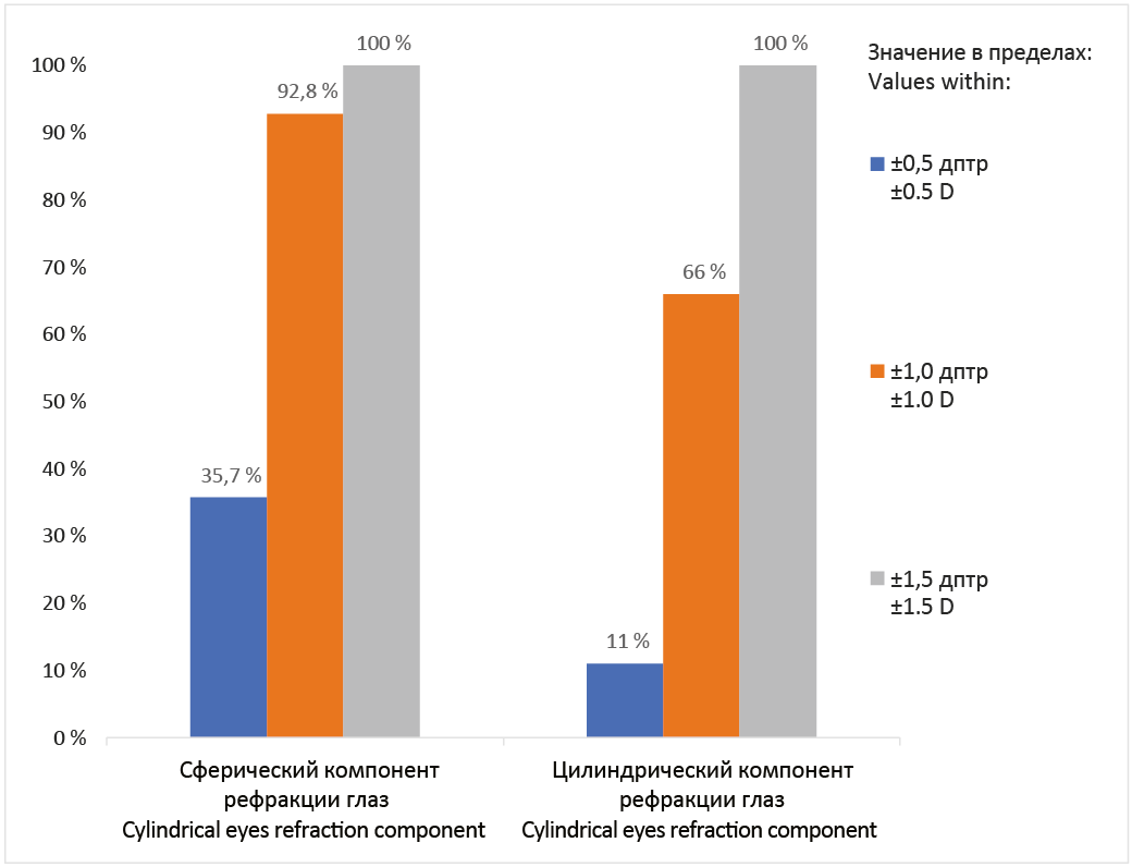
Relevance. In most countries of the world, applanation tonometers remain popular, such as the Maklakov tonometer and the Goldman tonometer. When measuring ophthalmotonus, such tonometers receive IOP values indirectly through the cornea, which certainly introduces its own errors into the measurement results. The creation of methods for measuring IOP different from the currently existing ones is relevant. Purpose: to create a new method for determining intraocular pressure without tonometry, based on the indicators of the individual profi le of the cornea with its parameters, indices and autorefractometry data. Materials and methods. Statistical analysis of 16 parameters of a keratotopograph (ALLEGRO Oculyzer, WaveLight Oculyzer II), data of an autorefractometer (TONOREF Nidek device) and data of tonometric intraocular pressure was carried out using a Maklakov tonometer (НГм2-«ОФТ-П») in 500 patients (1000 eyes). Among the sample population there were patients with both emmetropic refraction – 8 eyes (0.8%), and patients with refractive errors 992 eyes (99.2%), among them: 978 eyes (97.8%) had myopic refraction, 14 (1.4%) eyes had isolated refraction with myopic astigmatism. 889 eyes (88.9%) combined myopic refraction with myopic astigmatism. Results. Based on the analysis of keratotopographic parameters, autorefractometry values and tonometric IOP of 500 patients (1000 eyes), we have created a new method for determining intraocular pressure without tonometry, presented in the form of a mathematical model: Pt keratotopographic = 61.9 – 0.06 × SPH – 2.39 × Rf + 0.64 × Rmin – 0.15 × log2 (IVA) – 31.9 × CKI – 0.006 × Thickness. Conclusions. Based on the results obtained for determining IOP without physical interaction with the eye, the mathematical model created by us can be used in cases where the use of any tonometer is impossible.
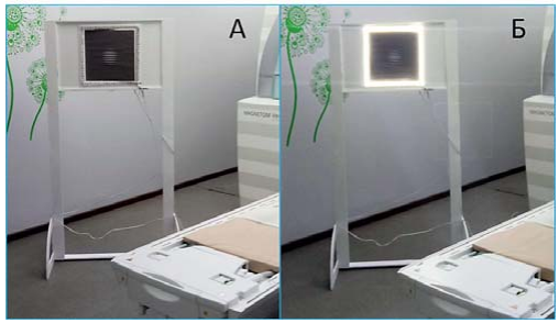
Background. In practical ophthalmology during cataract surgery, patients are increasingly being implanted with multifocal intraocular lenses (mfIOLs), which form several foci on the retina. The appearing of new factors creates conditions for human neuroadaptation, the parameters of which can be assessed using a modern research method – functional magnetic resonance imaging (fMRI). Purpose: to determine the change in the nature of the activation of the visual brain cortex in response to stimulation after surgery for the implantation of a multifocal artifi cial lens. Materials and methods. The study included patients with cataract (n = 22), who underwent structural neuroimaging and BOLD fMRI to assess neuronal activity before and after mfIOL implantation. The Gabor element was used as a stimulus for performing BOLD fMRI (unilluminated and ill uminated versions). fMRI data (positive BOLD effect) were processed using the SPM 12 software package in the Matlab R2017a environment. Evaluation of the research results was carried out at the individual and group levels. Results. When evaluating individual results after mfIOL implantation, patients show more local activation of the cerebral cortex, gravitating towards the area of the spur sulcus, while before surgery, the cortical response is more diffuse. In a group analysis in patients after mfIOL implantation, the total volume of the activation cluster of the cerebral cortex signifi cantly increased by 27 times. Maximum activation is noted in the pulvinar on the right and the lingual gyrus on the left. The activation of the cortex in the study with red illumination of Gabor element after surgery is less than in the study with white illumination, and less than in the study after surgery without illumination of Gabor element. Conclusions. A signifi cant but ambiguous change in the neuronal activity of the cerebral cortex on various stimulation options in patients after mfIOL implantation was established. Further work in this area is planned
Relevance. Some of the most current intraocular lenses (IOL) are lenses with an extended depth of focus. Lenses of this type allow you to reduce dependence on glasses by distributing light energy to long-range and focus at medium distance, without losing the quality of vision. Studies that analyze the results of implantation of this type of lens are few. Purpose: to evaluate the clinical outcomes of extended depth of focus (EDOF) intraocular lens (IOL) implantation. Materials and methods. Prospective randomized study enrolled 61 patients of 79 eyes (18 to 86 years) undergone uncomplicated phacoemulsifi cation with EDOF IOL Tecnis Symfony (USA) implantation. The study was conducted from November 2020 through November 2022. Uncorrected near visual acuity (UNVA); corrected near visual acuity (CNVA) and uncorrected and corrected distance visual acuity (UDVA and CDVA), monocular defocus curve and refractive outcomes were evaluated during a 3-month period. When calculating the IOL, the target spherical equivalent was –0.35 ± 0.11 D. Results. In the postoperative period 3 months, visual acuity were 0.7 or better in 82.4% UCDVA, 100% CDVA and 50% UCNVA. 3 months after surgery, UDVA and UNVA at 40 cm averaged 0.85 ± 0.15 and 0.69 ± 0.18, respectively. CDVA and CNVA at 40 cm was 0.98 ± 0.04 and 0.8 ± 0.14, respectively. A total of 85.7% of eyes achieved postoperative visual acuity about 0.5 for the range of defocus levels between +1.00 and −1.50 D. Conclusions. Cataract surgery with Tecnis Symfony EDOF IOL implantation provide functional levels of visual acuity in distance, intermediate zones. The near visual performance with this IOL might be signifi cantly enhanced using a micro-monovision approach
Orthokeratology has gained widespread clinical application in the last decade due to accumulated scientifi c data confi rming its effectiveness in inhibiting progressive myopia in children and adolescents. In cases of correction of other refractive errors, such as hypermetropia, mixed astigmatism, specialists inform patients about the possibilities of orthokeratology lenses (OKL) not often. This clinical case demonstrates the possibilities of customized, full-toric OKL to correct complex hyperopic astigmatism in an 11-year-old child. The patient was observed for several years with complex hyperopic astigmatism in ophthalmological clinics and followed the recommendations of specialists to use eyeglass correction. At the stage of transition from preschool to primary school, the child became shy of glasses and refused to wear them. In search of an alternative method of correction, parents sought advice in the special contact lens practice. The patient underwent a complete ophthalmological examination, including corneal topography. Taking into account the features of refraction and the shape of the cornea, the wishes of the child and parents, we have chosen customized orthokeratology lenses (OKL) with a fully toric design (full-toric) as method of correction. The OKL parameters were calculated by using the RGP Designer program. The lenses were made by OKVision Laboratory (Moscow). The fi tting of customized OKL allowed us to solve several tasks at once: the correction of complex hyperopic astigmatism and the freedom from using correction tools in the daytime. Despite the fact that orthokeratology is not the fi rst-line choice for optical correction of hyperopia, in some cases the method may become the only alternative to glasses and soft contact lenses
REVIEWS
Background. The increasing prevalence of progressive myopia and its complications necessitates the identifi cation of reliable diagnostic markers and new rational therapeutic tactics based on the results of studying the mechanisms of myopia development. Purpose: to assess the current understanding of the pathogenesis of progressive myopia in the light of the development of new effective methods of its control and treatment. Materials and methods. A bibliographic study of scientifi c publications of scientifi c information databases: Pubmed, eLibrary, Cyberleninka for the last 10 years was carried out. More than 100 sources were analyzed. The work included a review of 60 works. Results. This review considers both well-known and lesser-known theories that explain the causes and mechanisms of myopia progression. The publications demonstrate the importance of the problem of myopia development and allow us to signifi cantly expand the understanding of the mechanisms of development of this disease. The analysis of literature sources allowed us to provide important evidence of the role of heredity, s clera morphology, peripheral defocus and other factors in the development of progressive myopia, as well as the results of experimental studies on the key mechanisms of its development. However, the pathogenesis of progressive myopia is currently not fully understood. Moreover, each of the theories explaining the development of myopia has been repeatedly confi rmed in the results of other studies. This not only strengthens their evidence base, but also reveals common points where theories overlap, which gradually leads to the formation of a common consensus on the etiopathogenesis of this disease. Conclusions. Increased knowledge of the processes leading to the progression of myopia opens new opportunities for the development of effective methods that will ensure its control and stabilization, taking into account its pathogenesis.
WORKSHOP
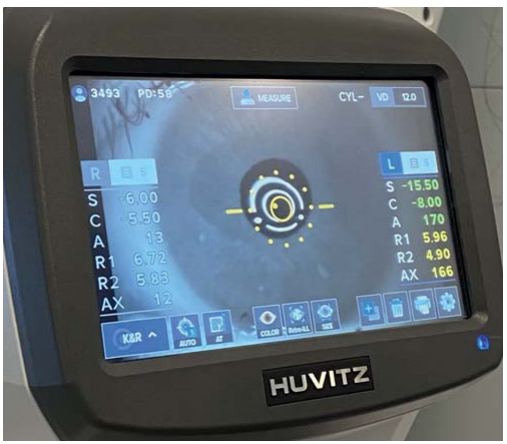
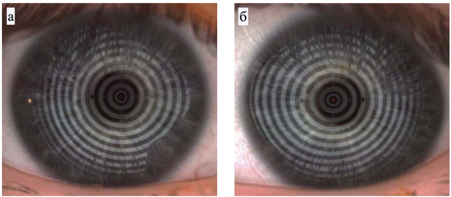
The article presents data on modern methods of diagnosis and monitoring of keratoconus. The main pathognomonic signs are described according to routine research methods: biomicroscopy, viso-, autoref- and keratometry, as well as special research methods such as keratotopography, keratotomography, optical coherence tomography, aberrometry, confocal microscopy and the study of biomechanical properties of the cornea. The “gold standard” in the diagnosis of subclinical keratoconus, today, is scanning keratotomography on a Scheimpfl ug camera with the Belin-Ambrósio enhanced ectasia protocol embedded in the device