


ORIGINAL ARTICLES
The increasing prevalence of myopia is observed all over the world, and Russia is no exception. In this regard, obtaining new data on its epidemiology among children and assessing methods of its control in real clinical practice are highly topical matters.
Purpose. The purpose of this work was to assess the current issues of the epidemiology and treatment of progressive myopia in children in various regions of the Russian Federation.
Materials and methods. We conducted a prospective multicenter epidemiological observational questionnaire study. This study involved 106 doctors from 53 regions of Russia and 2931 parents of myopic children.
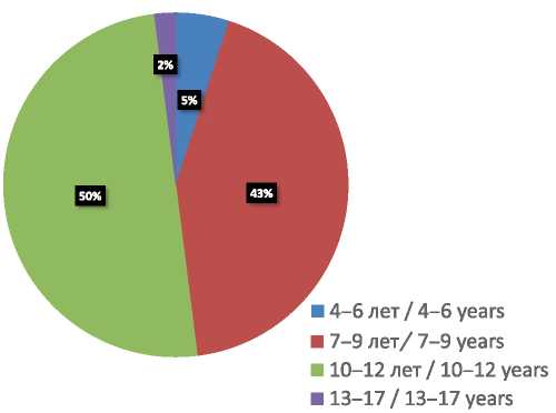
Results. 50% of the surveyed doctors noted that the manifestation of myopia is diagnosed in children aged 10–12 years, while 43% noted the same in children aged 7–9 years. According to 74.5% of doctors, the degree of the newly diagnosed myopia ranges from –1.25 to –3.00 diopters, 25.5% of doctors reported that it is below –1.0 diopters. The majority of doctors (73.6%) assess accommodation in myopic children, considering it one of the progression factors. 52.9% of ophthalmologists prescribe optical correction in cases when monocular distance visual acuity amounts to 0.6 (decimal scale) or lower values, while 29.2%, 16% and 1.9% of the surveyed ophthalmologists prescribe optical correction in cases when monocular distance visual acuity amounts to 0.7, 0.8 and 0.9 respectively.
The following optical methods for juvenile myopia control are recommended by the doctors: orthokeratology contact lenses (53.8%), spectacles for full correction (51.9%), peripheral defocus-inducing (bifocal) soft contact lenses (18.9%), while 4.7% of the surveyed doctors utilized other methods of myopia control, which were not indicated in the questionnaire.
Conclusion. In most cases, manifestation of myopia is diagnosed in children aged 7–12 years. Its degree ranges from –1.25 to –3.0 diopters, which indicates its late diagnosis; optical correction is prescribed mainly in cases when monocular distance visual acuity is 0.6 or lower; most ophthalmologists assess accommodation in myopic children, considering it a progression factor. As methods of myopia control, doctors utilize optical correction, device-assisted therapy and pharmacological treatment of accommodative disorders, while parents prefer methods that require minimum time expenditures.

Introduction. According to the studies, one out of three myopic patients with refraction greater than –6.00 D and an axial length greater than 26 mm is at high risk of facing low vision and loss of sight in the future. According to the results of medical examinations and screenings in carried out in Ivanovo, the prevalence of myopia in primary school children has increased three times during the past twenty years. Myopic children under 7 years old are six times more likely to have myopia progressed to higher degrees than children in which myopia onset took place later (at the age of 11–12 years). Optical interventions for myopia control such as orthokeratology and soft bifocal contact lenses have a strong body of evidence and are well accepted by ophthalmologists.
Purpose. The purpose of the present study was to investigate the effect of soft bifocal contact lenses on refraction, accommodation and axial length in children with progressive myopia.
Materials and methods. We observed 30 children aged 8–15 years with myopia progression rate of 0.82 D/year and accommodative weakness and instability. We prescribed OKVision PrimaBio Bi-focal design soft bifocal contact lenses (OKVision, Russia) that feature +4.00 D addition power on periphery. The effectiveness was estimated by monitoring refraction, accommodation and axial length every three months within a year.
Results. After 12 months of wearing soft bifocal contact lenses, the annual myopia progression rate decreased 4.3 times on average. We were able to stabilize myopia in 50% of the children during the period of monitoring. The use of this intervention had a strong effect on accommodation resulting in an increase of its amplitude and reserve.
Conclusion. The use of soft bifocal contact lenses has been proven to have a strong inhibitory effect on myopia progression rate. Myopia stabilization manifested itself as the absence of increase in myopic refraction and axial length as well as normalization of accommodative function.

Aim. To study the dynamics of changes in the values of higher order aberrations in amblyopia treatment and the correlation between higher-order aberrations and astigmatism in patients with hyperopic amblyopia.
Methods. This cohort prospective study included 36 patients (36 eyes) with refractive amblyopia aged 4 to 16 years. All patients had anisometropia: emmetropia in one eye and hyperopic astigmatism combined with refractive amblyopia of varying degrees in the other eye. Patients were divided into two groups depending on the degree of astigmatism. Astigmatism greater than 1.5 D was detected in 20 patients (55.5%) and astigmatism less than 1.5 D was detected in 16 patients (44.5%). All patients underwent a complex treatment, including twenty half-hour sessions of videocomputer autotraining using “Amblyotron” device during 20 days, in addition to constant wearing of glasses. Higher order aberrations were measured using the WaveScan Wavefront System aberrometer at the first visit and at 3-, 6 - and 12-month follow-up. A correlation analysis was performed to assess the relationship between higher order aberrations and astigmatism.
Results. There was a statistically significant difference in treatment success between groups with high and low astigmatism. In both groups, higher order aberrations were reduced during the treatment of amblyopia. When comparing the two groups, a significant difference in coma was found at 12-month follow-up (p = 0.043). At 12-month follow-up, coma showed a statistically significant correlation with astigmatism, and a stronger correlation with astigmatism was found in the group of patients with high astigmatism.
Conclusions. In patients with refractive amblyopia associated with astigmatism, the decrease in visual acuity is directly dependent on the values of higher-order aberrations, especially on the values of coma, which should be considered as the cause of the development of amblyopia.

Background. Optic neuritis is the first symptom of Davic’s disease in more than half of cases. Differential diagnosis of optic neuritis in the clinical practice is complicated due to the uniformity of the clinical pattern of inflammatory and demyelinating optic neuritis in the early stages of the disease. The approach to the management of patients with Davic’s disease is varied and requires a precise differentiation at the initial stages of its development.
Purpose. To determine early objective criteria for diagnosing the optic neuritis in the setting of Davic’s disease.
Materials and methods. We observed 31 patients (51 eyes), while the control group consisted of 12 healthy individuals (12 eyes). Research methods were both standard ophthalmic and specialized – optical coherence tomography, visual evoked potential test, magnetic resonance imaging of the brain and spinal cord.
Results. In patients with optic neuritis in the setting of Davic’s disease, optical coherence tomography revealed a smaller area of the optic nerve disc and neuroretinal belt as well as a decrease in macular volume and macular thickness. A reduction of the retinal ganglion cell complex and the inner plexiform layer was also revealed.
Conclusion. At the initial stages of Davic’s disease, it is necessary to conduct optical coherence tomography of the retina, perform visual evoked potential test as well as magnetic resonance imaging of the brain and spinal cord. Appropriate treatment at an early stage of the disease can reduce the rates of axonal degeneration and optic disc atrophy development.
REVIEWS
Ocular tuberculosis is a serious disease with a long recurrent course, often leading to a significant decrease in the visual functions and quality of life of patients as well as disability. In recent years, the incidence rate of the ocular tuberculosis in the Russian Federation has been declining: in 2016, according to a number of authors, its incidence amounted to 5.2%.
Purpose. To systematize literature data on the topic of ocular tuberculosis.
Materials and methods. We reviewed literature available on elibrary.ru, cyberleninka.ru websites and in “Clinical Ophthalmology” journal. Results. We suggested classification of the ocular tuberculosis lesions, analyzed modern diagnostic methods and treatment regimens and evaluated their effectiveness.
Conclusions. Literature review revealed that in order to improve the quality of detection, diagnosis and treatment of ocular tuberculosis, joint efforts of ophthalmologists and phthisiologists as well as use of modern methods of tuberculinodiagnosis are essential.
TECHNOLOGIES
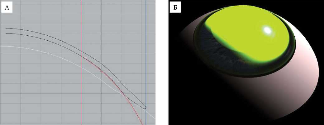
Corneal keratoectasia and dry eye syndrome (DES) are two of the most prevalent eye diseases. A number of studies is being carried out aimed at identifying and analyzing the annual increase in incidence of these diseases in population strata. Treatment and rehabilitation of patients with keratoectasia and DES remain topical issues in modern ophthalmology. Conventional treatment methods are not always effective, and scleral lenses (SCLs) may be considered an alternative. In this article, the authors expand on technology of manufacturing of OKVision® SMARTFIT™ scleral contact lenses.
DISCUSSION CLUB

LITERARE GUIDE
РЕЦЕНЗИИ

MEDICINE AND LAW
NEWS: WHAT? WHERE? WHEN?

ISSN 2686-8083 (Online)


